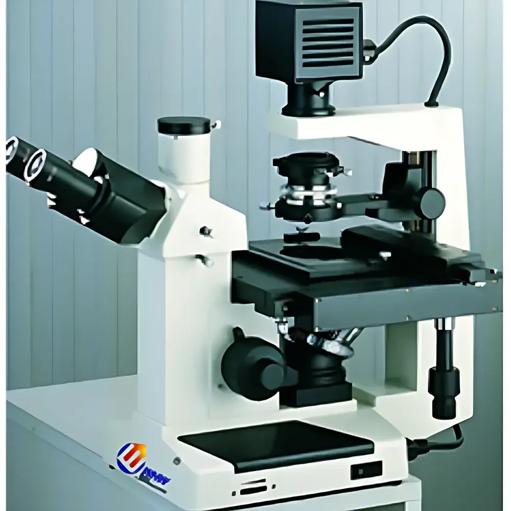
YANRUN BID-200 Inverted Biological Microscope
| Brand | YANRUN |
|---|---|
| Origin | Shanghai, China |
| Manufacturer Type | Direct Manufacturer |
| Product Category | Domestic (China-made) |
| Model | BID-200 Inverted Biological Microscope |
| Microscope Type | Inverted Microscope |
| Eyepiece Configuration | Binocular |
| Optical Magnification Range | 100×–400× |
| Mechanical Tube Length | 160 mm |
| Object Distance (Conjugate) | 195 mm |
| Eyepiece Port Diameter | 23.2 mm |
| Parfocal Distance | 10 mm |
| Standard Eyepiece | Wide-Field WF10× (Φ20 mm) |
| Objective Lenses | Long Working Distance Plan Achromatic PLL: 10×/0.25 (WD 8.8 mm), 25×/0.40 (WD 4.8 mm), 40×/0.60 (WD 3.3 mm) |
| Phase Contrast Objectives | PLL 10×/0.25 PHP2 |
| Trinocular Head | 30° Inclined, 100% Light Path for Imaging, Interpupillary Adjustment 53–75 mm |
| Focus Mechanism | Coaxial Coarse/Fine Focus with Adjustable Coarse Tension, Locking & Upper/Lower Limit Stops, Fine Focus Graduation: 2 µm |
| Nosepiece | 4-Position Revolving (Ball-Bearing Centered) |
| Stage | Dual-Layer Mechanical Stage (224 × 208 mm), Travel Range 130 × 79 mm, Removable Vernier Scale |
| Specimen Holders | Three Configurations — (1) 77.5 × 34 mm (for Ø68.5 mm round dishes), (2) 82 × 57 mm culture vessel support, (3) 129.5 × 86 mm (for Ø87.5 mm round dishes) |
| Transmitted Illumination | 6 V / 30 W Halogen Lamp with Continuous Brightness Control |
| Filters | Blue, Green, Ground Glass |
| Phase Contrast Assembly | Sliding-Type Annular Diaphragm System with Centering Telescope, 10× Phase Ring Plate, Adjustable Condenser (NA 0.4, WD 50 mm) |
| Condenser | NA 0.4, WD 55 mm, Integrated Phase Contrast Capability |
Overview
The YANRUN BID-200 Inverted Biological Microscope is an engineered optical platform designed specifically for live-cell observation, routine tissue culture monitoring, and long-term in vitro assays. Its inverted configuration places the objective lenses beneath the specimen stage—enabling direct access to cell culture vessels (e.g., Petri dishes, flasks, multi-well plates) without disturbing sample integrity or sterility. This architecture supports non-invasive, real-time visualization of adherent and suspension cells under physiological conditions. The system employs standard finite-conjugate optics (160 mm mechanical tube length) with parfocal, long-working-distance plan achromatic objectives—ensuring consistent focus across magnifications while maintaining ample clearance between objective front lens and culture vessel bottom. The optical path is optimized for high-contrast transmitted-light imaging, with integrated phase contrast capability for label-free visualization of unstained, transparent biological specimens.
Key Features
- Inverted optical design with fixed-stage configuration, supporting direct placement of standard cultureware including Ø68.5 mm, Ø87.5 mm round dishes, and rectangular T25/T75 flasks.
- Trinocular head inclined at 30°, offering full light-path allocation to imaging devices—compatible with C-mount adapters for USB/VIDEO digital cameras and photomicrographic systems.
- Four-position ball-bearing nosepiece ensures precise, repeatable objective alignment; optional five-position version available for expanded modularity.
- Dual-layer mechanical stage (224 × 208 mm) provides 130 × 79 mm travel range with vernier-scaled positioning—critical for systematic scanning of large-area cultures or multi-field time-lapse experiments.
- Coaxial coarse/fine focusing mechanism with adjustable coarse-tension control, mechanical lock, and dual-limit stops—enhancing operational stability during extended observation sessions.
- Integrated sliding-type phase contrast system with centering telescope, interchangeable annular plates (10× standard, optional 25×/40×), and height-adjustable condenser (NA 0.4, WD 50 mm)—supporting quantitative phase contrast alignment per ISO 10934-1 guidelines.
- 6 V / 30 W halogen illumination with continuous intensity regulation and standardized filter holders (blue, green, ground glass) for contrast optimization and reduced phototoxicity.
Sample Compatibility & Compliance
The BID-200 accommodates a broad range of commercially available cell culture formats—including standard 35 mm, 50 mm, and 60 mm Petri dishes; T25, T75, and T175 flasks; and multi-well plates (6–96-well). Customizable specimen holders (e.g., 77.5 × 34 mm, 129.5 × 86 mm, and 51.5 × 74.5 mm apertures) ensure mechanical stability and centration accuracy. All optical components comply with ISO 8578 (microscope nomenclature) and ISO 10934-1 (phase contrast performance standards). While not certified for GMP-regulated environments out-of-the-box, the instrument’s mechanical reproducibility, calibrated fine-focus graduation (2 µm), and traceable optical specifications support GLP-aligned documentation workflows when paired with validated digital imaging software.
Software & Data Management
The BID-200 serves as a hardware foundation for third-party imaging platforms via standard C-mount interface and USB/VIDEO output options. When integrated with compliant acquisition software (e.g., NIS-Elements, ZEN, or open-source MicroManager), it supports time-lapse capture, region-of-interest (ROI) tracking, and metadata embedding—including timestamp, magnification, objective ID, and illumination settings. Optional digital camera interfaces enable audit-trail generation compatible with FDA 21 CFR Part 11 requirements when deployed with electronic signature-enabled software suites. Raw image data is stored in vendor-neutral TIFF or OME-TIFF formats to ensure long-term archival integrity and cross-platform interoperability.
Applications
- Real-time monitoring of cell proliferation, migration, and morphology changes in monolayer and co-culture systems.
- Phase contrast imaging of unstained primary neurons, fibroblasts, stem cells, and organoid cultures—eliminating fixation and staining artifacts.
- Quality control of bioprocesses including transfection efficiency assessment, confluency estimation, and contamination screening in upstream biomanufacturing.
- Support for basic fluorescence add-ons (with external epi-illumination modules) in teaching laboratories and preclinical research settings.
- Long-duration live-cell imaging experiments requiring mechanical stability, thermal consistency, and minimal operator intervention.
FAQ
What culture vessels are directly supported without adapter modification?
Standard Ø68.5 mm and Ø87.5 mm round Petri dishes, as well as 82 × 57 mm rectangular culture vessels, are natively accommodated using the included specimen holders.
Is the microscope compatible with digital imaging systems?
Yes—the trinocular port supports C-mount adapters, and USB/VIDEO output interfaces are available as factory options for integration with CMOS/CCD cameras.
Can phase contrast be used with all objective magnifications?
The standard configuration includes a 10× phase ring plate. Optional 25× and 40× phase rings, along with corresponding PLL PHP2 objectives, are available for expanded phase contrast capability.
Does the system meet international optical calibration standards?
All optical parameters—including parfocality (10 mm), tube length (160 mm), and objective conjugate distance (195 mm)—adhere to ISO 8578 and ISO 10934-1 specifications for inverted microscopes.
What maintenance protocols are recommended for long-term optical fidelity?
Routine cleaning of optical surfaces with lens-grade solvents, annual verification of phase ring alignment using the included centering telescope, and periodic recalibration of fine-focus graduation are advised per manufacturer service guidelines.
Related Products


