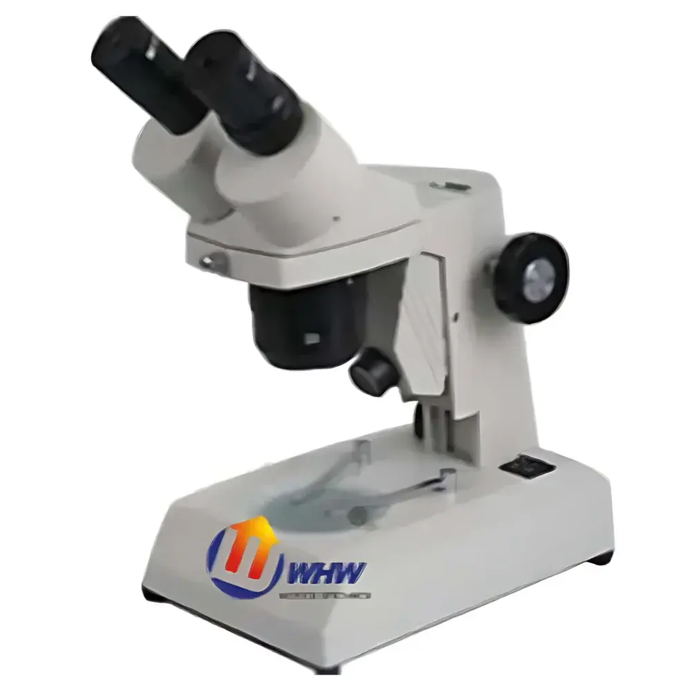
YANRUN PXS-2040 Advanced Stereo Microscope
| Brand | YANRUN |
|---|---|
| Origin | Shanghai, China |
| Manufacturer Type | Original Equipment Manufacturer (OEM) |
| Model | PXS-2040 |
| Total Magnification Range | 10×–30× (standard configuration) |
| Working Distance | 90 mm |
| Objective Lenses | 1× and 3× (interchangeable) |
| Illumination | 6 V / 20 W halogen top light |
| optional bottom illumination | 6 V / 20 W incandescent, 5 W fluorescent, or 1 W LED |
Overview
The YANRUN PXS-2040 Advanced Stereo Microscope is a high-stability, dual-port stereo inspection system engineered for precision visual analysis in industrial quality control, electronics assembly, materials science laboratories, and educational settings. Utilizing Greenough optical design principles, the PXS-2040 delivers true stereoscopic imaging with independent optical paths—ensuring natural depth perception, minimal parallax error, and consistent resolution across the entire field of view. Its fixed 1× and 3× objective pair provides a continuous magnification range of 10×–30× when paired with standard WF10× wide-field eyepieces, extendable to 40× via optional WF15× or WF20× oculars. Designed for ergonomic operation over extended periods, the microscope features a 45° inclined, 360° rotatable binocular head with interpupillary adjustment (55–75 mm), enabling optimal user alignment and reduced ocular fatigue during routine inspection tasks.
Key Features
- Robust mechanical architecture with gear-and-rack coarse focusing mechanism, incorporating dual-stage handwheel tension control and built-in upper/lower travel limit stops for repeatable Z-axis positioning.
- Fixed-focus objective turret supporting rapid switching between 1× and 3× objectives—eliminating focus drift during magnification changes and enhancing measurement reproducibility.
- Extended 90 mm working distance accommodates bulky samples, manipulators, soldering tools, and micro-probing fixtures without obstruction—ideal for PCB rework, metallurgical sample preparation, and biological dissection workflows.
- Modular illumination system: integrated 6 V / 20 W halogen top illuminator ensures uniform, color-stable incident lighting; optional transmitted-light modules include 6 V / 20 W incandescent, 5 W fluorescent, and energy-efficient 1 W LED base lights—each compatible with standard Koehler illumination alignment protocols.
- Dual-sample stage configuration: includes interchangeable 75 mm diameter black/white contrast plate and ground-glass diffuser plate—supporting both high-contrast surface inspection and low-glare translucent specimen observation.
Sample Compatibility & Compliance
The PXS-2040 supports broad sample dimensional compatibility—from small electronic components (SMD resistors, QFN packages) to irregularly shaped metallographic mounts, botanical specimens, and polymer test coupons. Its open-stage geometry and unobstructed optical path meet ISO 8549-1:2021 requirements for stereo microscope mechanical design and dimensional stability. While not certified to ISO/IEC 17025 as a calibrated metrology instrument, the system’s mechanical repeatability and optical consistency align with GLP-compliant documentation practices for non-quantitative visual inspection procedures in QC/QA environments. All electrical components comply with IEC 61010-1:2012 safety standards for laboratory equipment.
Software & Data Management
The PXS-2040 operates as a standalone optical platform with no proprietary firmware or embedded software. However, it is fully compatible with third-party digital imaging solutions—including USB3.0 CMOS cameras (e.g., AmScope MU1403, OMAX 10MP models) and industry-standard acquisition software such as ToupView, Motic Images Plus, or ImageJ. When coupled with calibrated stage micrometers and NIST-traceable reference standards, image capture workflows support audit-ready documentation per FDA 21 CFR Part 11 requirements—provided users implement external electronic signature controls and secure data storage protocols. Raw image metadata (magnification, illumination mode, date/time stamp) can be embedded via EXIF tagging in TIFF or PNG formats.
Applications
- Electronics manufacturing: solder joint inspection, component placement verification, counterfeit IC detection, and post-reflow defect mapping.
- Materials testing labs: fracture surface analysis, coating thickness estimation (via edge profiling), inclusion identification in cast alloys, and fiber orientation assessment in composites.
- Educational microscopy: comparative anatomy studies, mineral identification, entomological specimen examination, and introductory metallurgy demonstrations.
- Forensic document analysis: ink differentiation, paper fiber structure evaluation, and latent mark enhancement under variable contrast illumination.
- Jewelry and precision machining: surface finish grading, tool wear monitoring, and micro-defect triage prior to SEM analysis.
FAQ
Is the PXS-2040 suitable for photomicrography?
Yes—the trinocular port (optional accessory) enables simultaneous visual observation and high-resolution digital imaging using C-mount adapters compatible with most scientific-grade CCD/CMOS sensors.
Can the working distance be extended beyond 90 mm?
No—the optical tube length and objective focal plane are mechanically fixed; however, auxiliary long-working-distance auxiliary lenses (e.g., 0.5× or 2× Barlow systems) may be mounted externally to modify effective WD and magnification—subject to resolution trade-offs.
Does the system support fluorescence observation?
Not natively—the halogen and incandescent sources lack excitation bands required for fluorescence; however, third-party LED-based epi-illumination modules with 365 nm or 470 nm emission can be retrofitted with appropriate filter cubes.
What maintenance is recommended for long-term optical performance?
Annual cleaning of prism surfaces with lens-grade solvent and lint-free wipes; biannual gear lubrication using silicone-based grease; and periodic verification of interpupillary alignment and diopter balance per ISO 8549-2:2019 guidelines.


