Yokogawa FlowCam Nano Oil-Immersion Flow Imaging Microscope for Nanoparticle Characterization
| Brand | Yokogawa Fluid Imaging Technologies |
|---|---|
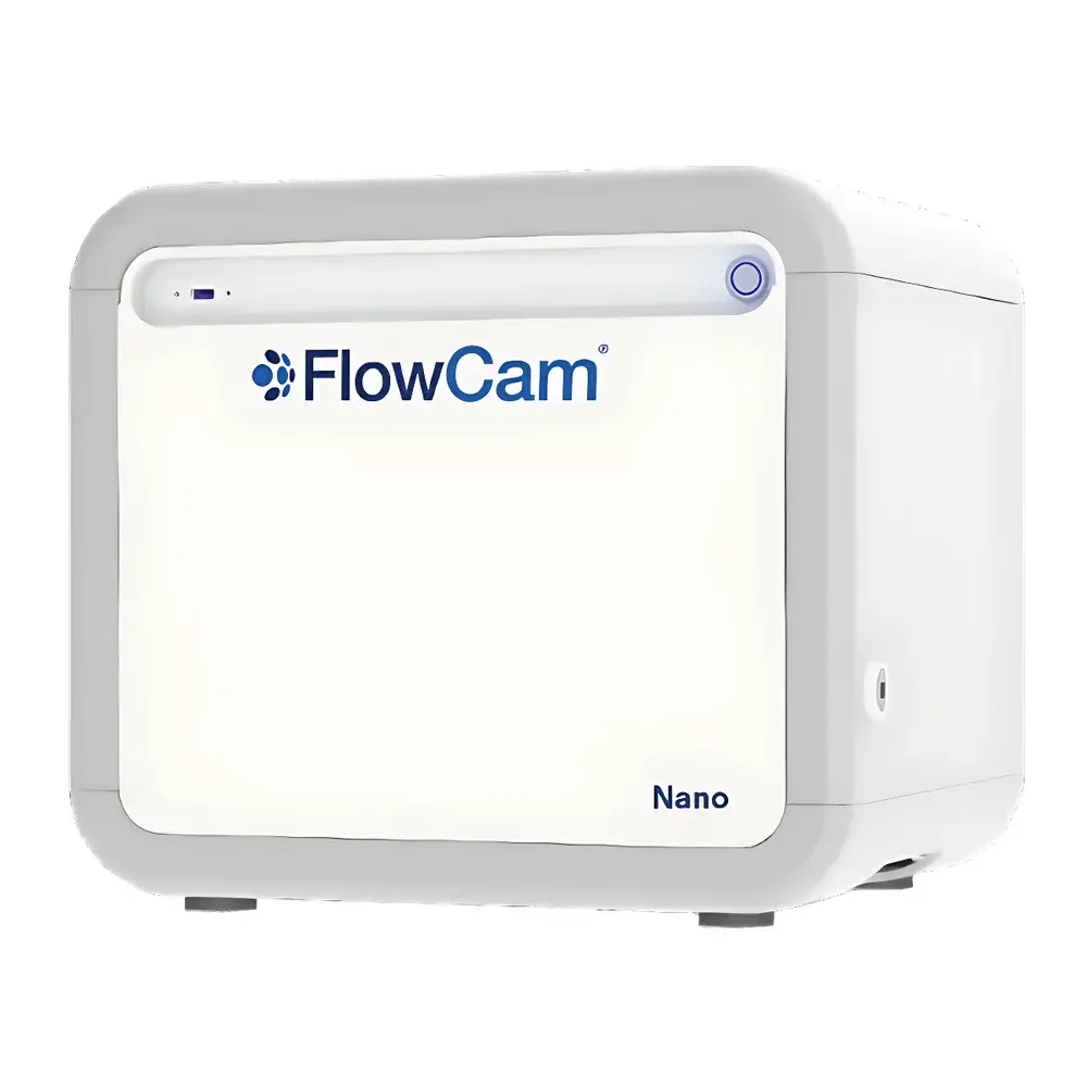
| Model | FlowCam Nano |
| Detection Range | 100 nm – 2 µm |
| Objective Magnification | 40× (oil-immersion) |
| Flow Cell Depth | 60 µm |
| Minimum Sample Volume | 50 µL |
| Flow Rate | 25 µL/min |
| Camera Resolution | 1440 × 1080 pixels (monochrome CMOS) |
| Frame Rate | up to 130 fps |
| Autofocus | Fully automated |
| Software | VisualSpreadsheet® with 21 CFR Part 11 compliance option |
| Dimensions | 44 cm (W) × 36 cm (D) × 39 cm (H) |
| Weight | 23 kg (operational), 45 kg (shipping) |
| Power | 100–240 VAC, 50/60 Hz, max. 92 W |
Overview
The Yokogawa FlowCam Nano is an oil-immersion flow imaging microscope engineered for high-fidelity visualization and quantitative morphological analysis of nanoparticles in suspension. Unlike conventional light microscopy or ensemble-based techniques (e.g., DLS or laser diffraction), the FlowCam Nano employs dynamic flow imaging—capturing individual particle images in real time as they pass through a precisely defined optical field within a microfluidic flow cell. Its core innovation lies in the use of immersion oil matched to the refractive index of the carrier fluid, eliminating spherical aberration at high magnification (40×) and enabling diffraction-limited resolution down to 100 nm. This allows unambiguous discrimination between primary nanoparticles, submicron aggregates, and co-flowing contaminants—critical for biopharmaceutical formulation development, nanomaterial safety assessment, and regulatory-compliant particulate analysis per USP , ISO 21501-4, and ICH Q5A(R2). The system operates without sample dilution, labeling, or centrifugation, preserving native particle state and enabling direct correlation of size, shape, transparency, and spatial distribution.
Key Features
- Patented oil-immersion optical path optimized for aqueous and organic suspensions, delivering consistent sub-200 nm resolution across the full 100 nm – 2 µm detection range
- Automated, closed-loop autofocus system ensuring pixel-level focus stability across multi-hour acquisition runs and variable sample viscosities
- High-speed monochrome CMOS camera (1440 × 1080 px, ≥130 fps) synchronized with micro-injection pumping (25 µL/min flow rate) for statistically robust particle sampling
- Integrated 60 µm-deep flow cell with laminar flow profile, minimizing shear-induced aggregation and enabling accurate hydrodynamic diameter estimation
- Real-time image segmentation and extraction—no post-acquisition thresholding artifacts; each particle image is saved losslessly with full metadata (timestamp, position, flow velocity)
- Compliance-ready architecture: optional 21 CFR Part 11 software package with electronic signatures, audit trail, and role-based access control for GLP/GMP environments
Sample Compatibility & Compliance
The FlowCam Nano accommodates a broad spectrum of liquid-phase samples—including protein therapeutics, lipid nanoparticles, polymer dispersions, colloidal silica, vaccine adjuvants, and environmental aqueous extracts—without requiring derivatization or fixation. Its non-destructive, label-free operation preserves biological activity and aggregation kinetics. Regulatory alignment includes support for USP , , and method validation protocols, particularly where orthogonal particle characterization is mandated for parenteral product release. The system meets ISO 21501-4 requirements for calibration traceability using NIST-traceable polystyrene standards and supports IQ/OQ documentation packages compliant with FDA and EMA expectations. All hardware components are CE-marked and RoHS-compliant; software validation kits include test scripts, failure mode logs, and raw data integrity verification tools.
Software & Data Management
VisualSpreadsheet® serves as the analytical engine—combining acquisition control, real-time image processing, and multidimensional statistical reporting in a single interface. It computes over 40 morphological descriptors per particle (e.g., circularity, convexity, aspect ratio, edge gradient, intensity variance, bio-volume approximations), enabling classification by structural signature rather than size alone. Users define custom gates using Boolean logic across any parameter combination, then export filtered populations to CSV, Excel, or HDF5 for downstream PCA, clustering, or machine learning workflows. Batch processing supports comparative analysis across multiple runs, including temporal stability studies or lot-to-lot QC comparisons. Audit trails record every user action, parameter change, and data export event with timestamp and operator ID. Optional cloud synchronization enables secure remote review and collaborative annotation across global R&D teams.
Applications
- Biopharmaceutical Development: Quantifying subvisible protein aggregates, silicone oil droplets, glass delamination fragments, and cellulose fibers in mAb formulations; supporting forced degradation studies and container-closure compatibility assessments
- Nanomedicine Characterization: Resolving liposomal polydispersity, PEGylated nanoparticle coating uniformity, and extracellular vesicle morphology in exosome therapeutics
- Environmental Monitoring: Identifying and enumerating microplastics (<500 nm), cyanobacterial colonies, diatom frustules, and algal bloom species in freshwater, seawater, and wastewater matrices
- Materials Science: Assessing carbon black dispersion quality, CMP slurry particle integrity, toner pigment agglomeration, and ceramic powder sintering precursor homogeneity
- Food & Beverage QA: Detecting starch granule gelatinization, yeast viability morphology, pollen contamination, and flavor encapsulant rupture in emulsions
- Quality Control: Validating filter integrity, monitoring cleaning validation residues, and verifying batch consistency in sterile fill-finish operations
FAQ
What is the smallest particle size reliably resolved by the FlowCam Nano?
The system achieves consistent visualization and measurement of particles ≥100 nm under standard aqueous conditions using 40× oil-immersion optics and calibrated illumination. Resolution is verified per ISO 13322-2 using certified latex sphere standards.
Can the FlowCam Nano differentiate between amorphous aggregates and crystalline precipitates?
Yes—through combined analysis of transparency (optical density), edge sharpness (perimeter roughness), and internal texture (intensity variance), users can distinguish translucent protein aggregates from highly refractive crystalline salts or metal oxides.
Is method transfer supported between FlowCam models?
VisualSpreadsheet® maintains backward-compatible project files and parameter sets. Migration from FlowCam 5000 or 8000 to Nano requires recalibration of size scaling and focus offset but retains all classification logic and report templates.
How does the system handle viscous or particulate-laden samples?
The micro-pumping system (250 µL syringe volume) supports programmable ramped flow rates and pressure monitoring. Clogging mitigation includes automatic flow reversal, ultrasonic cell cleaning cycles, and optional inline filtration (0.45 µm) pre-injection.
Does Yokogawa provide application-specific validation support?
Yes—dedicated application scientists deliver protocol development, method qualification (precision, accuracy, LOD/LOQ), and regulatory submission support aligned with ICH, USP, and regional health authority expectations.
")


