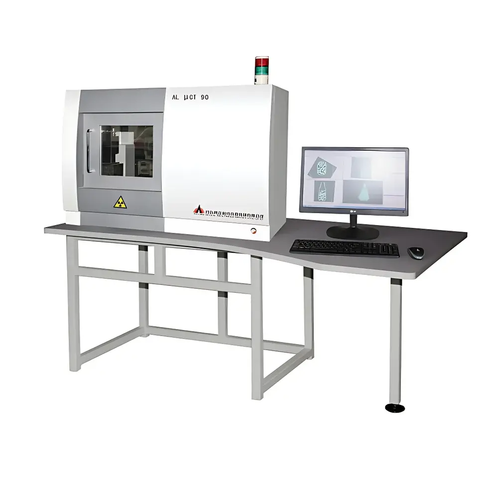
AoLong aolong Lab-Specific In Vivo Micro-CT Imaging System
| Brand | AoLong |
|---|---|
| Origin | Liaoning, China |
| Manufacturer Type | Direct Manufacturer |
| Product Category | Domestic |
| Model | Lab-Specific In Vivo Micro-CT Imaging System |
| Instrument Type | Tomographic Imaging |
| Tube Voltage | 20–90 kV |
| Focal Spot Size | 5 µm |
| Spatial Resolution | 3 µm |
| Density Resolution | 0.3–0.5% |
| Scan Geometry | Cone-Beam |
| Detector | Digital Flat-Panel Detector |
| Scan Time | 30 s – 2 min |
| Image Reconstruction Speed | 1024×1024, 720 projections |
Overview
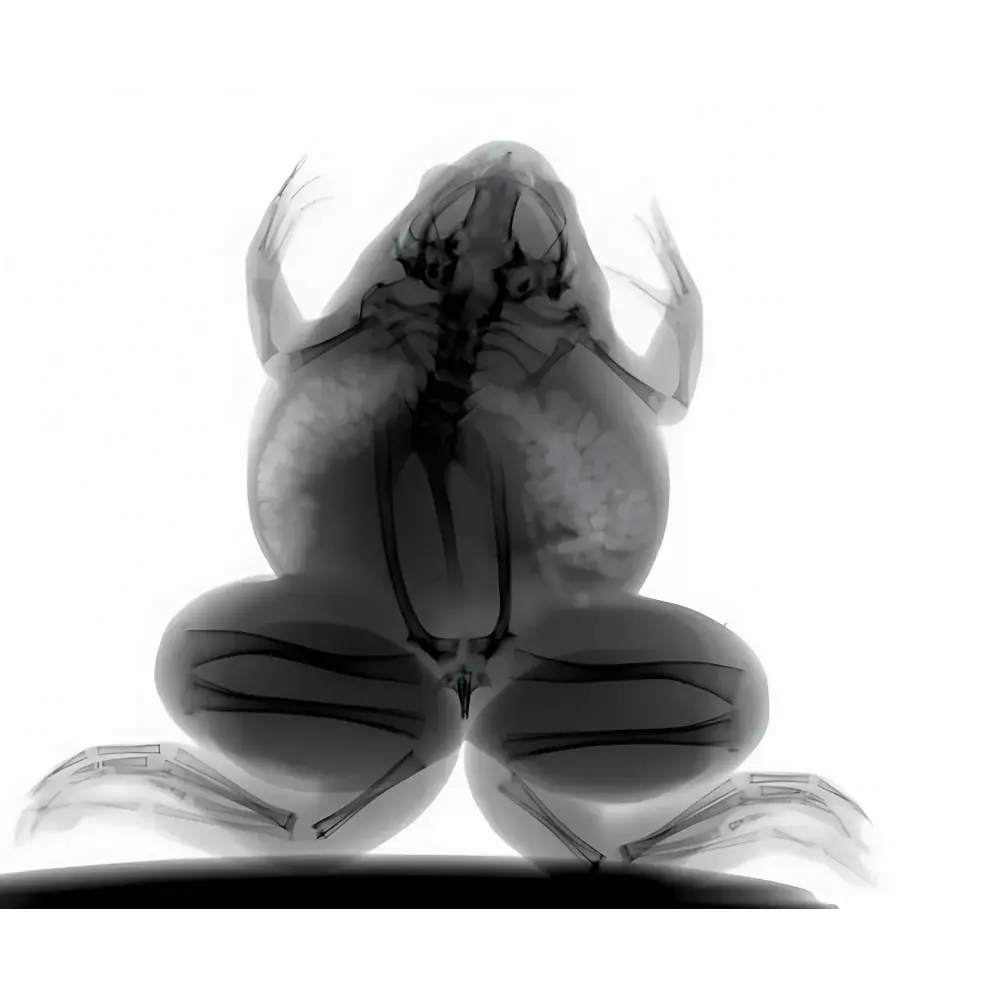
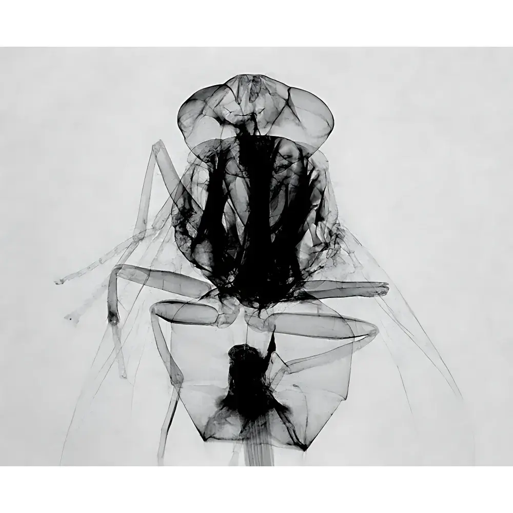
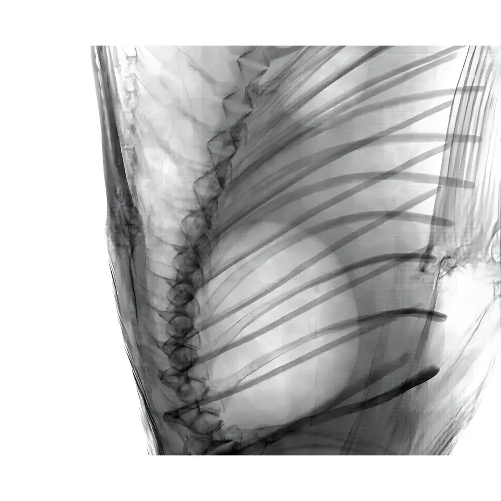
The AoLong Lab-Specific In Vivo Micro-CT Imaging System is a benchtop preclinical computed tomography platform engineered for non-invasive, high-resolution three-dimensional structural imaging of live small animals and biological specimens. Based on cone-beam X-ray microtomography (CBCT), the system delivers quantitative volumetric data without requiring tissue sectioning or contrast agent administration in many applications. It operates across a tunable tube voltage range (20–90 kV), enabling optimization for diverse sample densities—from soft tissues to mineralized structures—while maintaining sub-3-µm spatial resolution under optimal acquisition conditions. The system is purpose-built for longitudinal studies in pharmacology, oncology, orthopedics, and developmental biology, where repeated in vivo measurements must preserve animal viability and experimental integrity.
Key Features
- Sub-micron focal spot X-ray source (5 µm) ensuring minimal geometric blur and high-fidelity projection data.
- Digital flat-panel detector with high dynamic range and low electronic noise, supporting both fast real-time preview and high-SNR acquisition modes.
- Cone-beam geometry optimized for rapid volumetric coverage—full 360° rotation scans completed in as little as 30 seconds, with full-volume reconstruction delivered in under 2 minutes.
- Real-time image reconstruction engine: capable of reconstructing a 1024×1024 slice from 720 projections in 0.2 seconds per slice, enabling immediate post-scan qualitative assessment and iterative protocol refinement.
- Integrated thermal management and mechanical stabilization to minimize drift during multi-hour longitudinal sessions.
- Modular gantry design accommodating adjustable animal positioning stages, physiological monitoring interfaces (optional), and respiratory gating inputs for motion-compensated imaging.
Sample Compatibility & Compliance
The system supports live rodent models (mice, rats), zebrafish embryos, ex vivo tissue samples, and small botanical specimens. Anesthesia-compatible animal holders with integrated temperature and respiration monitoring ports ensure compliance with institutional animal care and use committee (IACUC) standards. All hardware and firmware meet CE marking requirements for laboratory equipment (2014/30/EU EMC Directive and 2014/35/EU Low Voltage Directive). Data handling workflows support audit-trail generation and user-access logging, facilitating alignment with GLP-compliant study documentation practices. While not FDA-cleared for clinical diagnostics, the system adheres to ISO 13485–aligned quality control protocols during manufacturing and calibration.
Software & Data Management
Bundled with AoLong MicroView™ Acquisition & Reconstruction Suite (v5.x), the system provides DICOM-compliant image export, NIfTI support, and native integration with open-source analysis platforms including 3D Slicer and ITK-SNAP. Volume rendering, region-of-interest (ROI) segmentation, bone morphometry (BMD/BV/TV), and dynamic density thresholding are implemented via validated algorithms traceable to ASTM E1255 and ISO 12706 standards. Raw projection datasets and reconstructed volumes are stored with embedded metadata (acquisition parameters, timestamp, operator ID), satisfying 21 CFR Part 11 readiness when deployed with network-authenticated user accounts and encrypted storage configurations.
Applications
- Longitudinal tumor growth monitoring and anti-angiogenic therapy response assessment.
- Quantitative osteoporosis modeling: trabecular architecture analysis, cortical thickness mapping, and fracture risk simulation.
- Pulmonary airway remodeling in asthma and COPD murine models.
- Vascular casting and micro-angiography using iodinated contrast agents.
- Developmental phenotyping in transgenic zebrafish and mouse embryos.
- Ex vivo validation of histological findings through co-registered virtual sectioning.
FAQ
Is this system suitable for longitudinal imaging of the same animal over multiple time points?
Yes—the system supports repeat scanning with consistent positioning accuracy (<±15 µm) and dose-controlled exposure protocols to minimize cumulative radiation impact.
What is the minimum detectable density difference under standard acquisition settings?
Density resolution is specified at 0.3–0.5% for 10-mm-thick water-equivalent phantoms at 70 kV and 100 µA, per IEC 61223-3-5 test methodology.
Can the system interface with third-party physiological monitoring devices?
Yes—via TTL-triggered synchronization ports and analog input channels compatible with industry-standard pulse oximeters, ECG amplifiers, and ventilators.
Does the software support automated batch reconstruction and scripting?
MicroView™ includes Python API access and CLI mode for unattended processing pipelines, including dose-optimized parameter inheritance across session templates.
Is service and calibration support available outside mainland China?
AoLong maintains authorized service partners in Germany, South Korea, and Brazil; remote diagnostics and annual calibration verification are supported via secure VPN-enabled maintenance mode.


