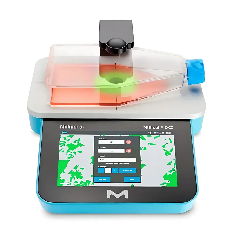
Millicell® DCI Digital Cell Imager
| Brand | Millipore |
|---|---|
| Origin | USA |
| Model | MDCI10000 |
| Dimensions (L×W×H) | 6 in × 11 in × 12 in |
| Weight | 5.67 lb |
| Connectivity | USB / Ethernet / Wi-Fi |
| Sample Compatibility | Adherent cells, microtissues, organoids |
| Data Management | Web-based cloud platform with audit-trail-capable image archiving and time-series growth analytics |
| Regulatory Context | Designed to support GLP-compliant cell culture monitoring |
Overview
The Millicell® DCI Digital Cell Imager is a benchtop, non-invasive imaging system engineered for quantitative, label-free assessment of adherent mammalian cell cultures in standard multiwell plates, flasks, and Petri dishes. Unlike traditional endpoint assays requiring trypsinization or staining, the DCI employs high-resolution brightfield microscopy coupled with real-time image acquisition and AI-assisted morphometric analysis to deliver objective measurements of confluence, cell count, and morphological parameters—including area coverage, nuclear-to-cytoplasmic ratio approximations, and colony dispersion metrics. Its optical architecture is optimized for phase-contrast–like contrast enhancement without external optics or sample manipulation, enabling repeated longitudinal monitoring of the same culture vessel over days or weeks. This capability supports rigorous experimental reproducibility, reduces biological variability introduced by passaging, and preserves precious primary or low-passage cell stocks for downstream functional assays.
Key Features
- Label-free, non-destructive imaging: No enzymatic detachment, fixation, or fluorescent labeling required—maintains cellular integrity and viability throughout measurement cycles.
- Automated confluence quantification: Algorithm-driven pixel-intensity segmentation calibrated against standardized reference cultures (e.g., NIH/3T3, HEK293, iPSC-derived lineages) yields ±3% inter-run CV for confluence estimates across 0–100% range.
- Integrated cell counting engine: Differentiates viable adherent cells from debris or confluent monolayers using adaptive thresholding and shape-based clustering—validated against hemocytometer counts for suspension-adapted lines post-detachment.
- Modular hardware interface: Supports USB 3.0 direct connection for local operation or Ethernet/Wi-Fi integration into institutional LIMS or ELN ecosystems with TLS 1.2–secured data transmission.
- User-configurable acquisition protocols: Adjustable focus depth, exposure time, and field-of-view tiling per well—enabling consistent imaging across 6-, 24-, 48-, and 96-well formats.
- On-device processing: All image analysis occurs locally on the embedded ARM-based controller; raw TIFFs and annotated JPEGs are exported with EXIF metadata including timestamp, objective magnification, and environmental sensor logs (ambient temperature/humidity if integrated).
Sample Compatibility & Compliance
The DCI is validated for use with standard tissue-culture-treated polystyrene surfaces (e.g., Corning Costar®, Falcon®), as well as hydrogel-coated or ECM-functionalized substrates used in 3D microtissue and organoid culture. It accommodates transparent-bottom vessels up to 22 mm height and supports imaging of heterogeneous populations—including co-cultures of fibroblasts and epithelial cells—without spectral interference. The system complies with IEC 61000-6-3 (EMC emissions) and IEC 61000-6-2 (immunity), and its software architecture supports audit trail generation, user access control, and electronic signature workflows aligned with FDA 21 CFR Part 11 requirements when deployed with validated cloud infrastructure. Routine performance verification follows ISO 21501-4 (particle size distribution via image analysis) principles adapted for cellular objects.
Software & Data Management
The DCI operates via a responsive web application accessible from any modern browser (Chrome, Edge, Firefox) without client-side installation. Image datasets are stored in encrypted AES-256 containers within a FIPS 140-2–certified cloud repository, with version-controlled metadata tagging (e.g., passage number, media batch ID, operator ID). Time-series confluence curves export to CSV or MATLAB .mat format; all images retain embedded DICOM-SR–compatible structured reporting fields for integration into PACS-adjacent research informatics platforms. Role-based permissions (admin, analyst, reviewer) enforce separation of duties; session logs record every image capture, parameter modification, and report generation event with ISO 8601 timestamps and IP geolocation.
Applications
- Standardization of seeding density in CRISPR editing workflows to minimize clonal heterogeneity.
- Longitudinal monitoring of iPSC differentiation efficiency across neural rosette formation timelines.
- QC release testing for biomanufacturing cell banks—tracking doubling time consistency across master/working cell stocks.
- Drug cytotoxicity screening using real-time confluence loss kinetics (IC50 derivation without LDH/MTS endpoint bias).
- Validation of scaffold-based 3D tumor spheroid growth under hypoxic gradients in perfusion bioreactors.
- Teaching laboratories: Objective assessment of student-performed transfection efficiency via GFP+ cell area expansion rates.
FAQ
Does the DCI require calibration with reference beads or cell standards?
Yes—annual factory recalibration is recommended, and users must perform daily verification using the included NIST-traceable polystyrene microsphere slide (10 µm nominal diameter) to confirm optical resolution and pixel scaling accuracy.
Can the system distinguish between live and dead adherent cells without staining?
No—viability discrimination requires membrane-impermeant dyes (e.g., propidium iodide) or metabolic probes; however, morphology-based anomaly detection (e.g., blebbing, shrinkage) can flag potential apoptosis events for targeted follow-up.
Is offline operation supported for secure environments with no internet access?
Yes—the onboard Linux OS enables full image acquisition, analysis, and local file export via USB drive; cloud sync resumes automatically upon network restoration without data loss.
What is the maximum frame rate for time-lapse imaging?
Up to 1 frame per minute across 96 wells (with region-of-interest cropping); continuous acquisition at full resolution is limited to single-well mode at 0.5 fps.
How does the DCI handle edge effects in low-confluence wells?
The software applies adaptive vignetting correction and peripheral masking based on vessel geometry recognition—validated down to 2% confluence with <5% relative error versus manual ground-truth annotation.


