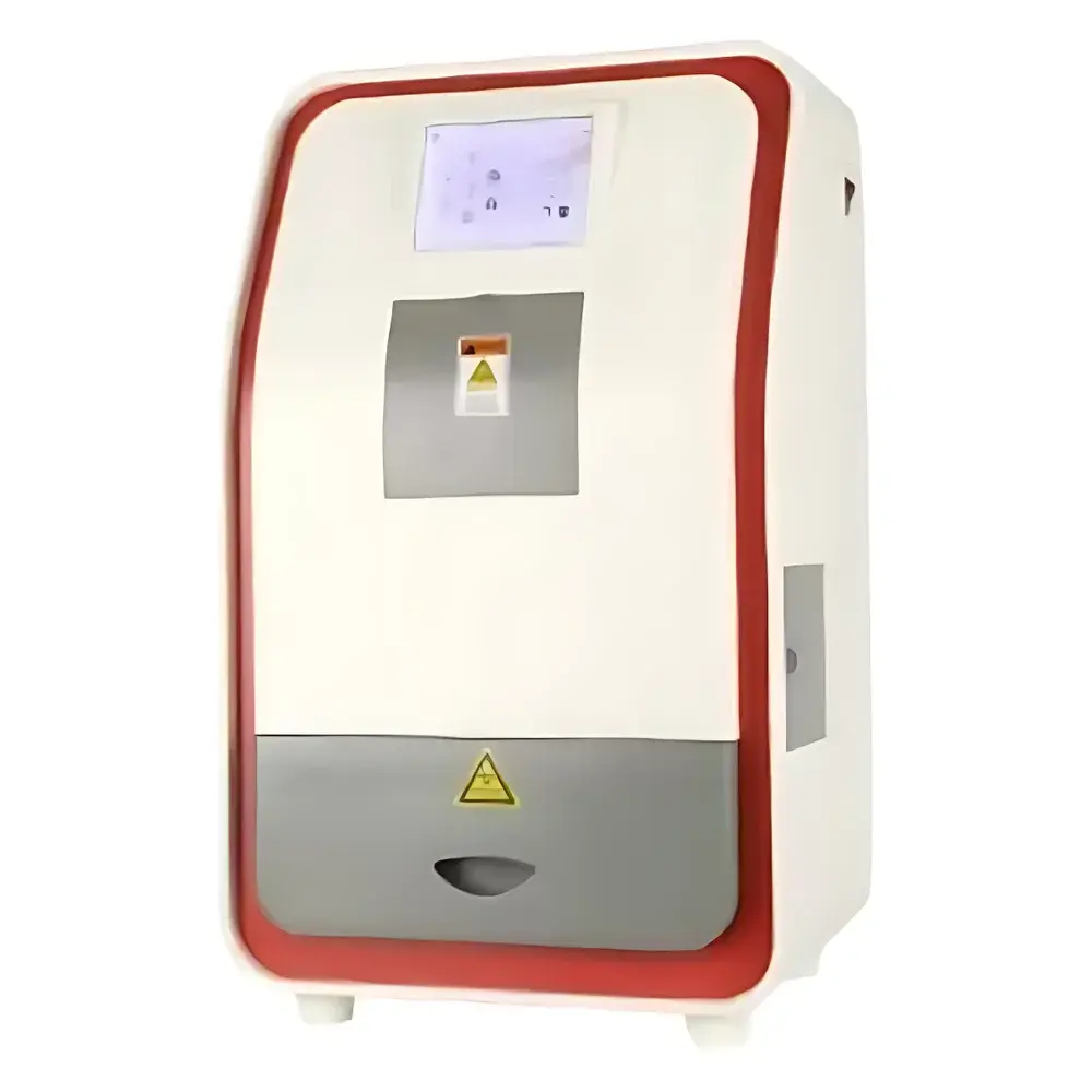

Jipeng JP-2880Plus Gel Imaging System
| Brand | Jipeng |
|---|---|
| Origin | Shanghai, China |
| Model | JP-2880Plus |
| Instrument Type | Standard Gel Imaging System |
| CCD Resolution | 3096(H) × 2080(V), 6.4 Megapixels |
| Bit Depth | 16-bit |
| Dynamic Range | 4.5 OD |
| CCD Sensor Size | 1/2.5" |
| Detection Sensitivity | ≤ 0.01 ng double-stranded DNA stained with ethidium bromide (EB) |
| Signal-to-Noise Ratio | ≥ 56 dB |
| Lens | Motorized optical zoom lens, 8–48 mm, 6× zoom, f/1.2 aperture |
| Transmission UV Source | 302 nm (standard), optional 254 nm / 365 nm reflectance UV |
| Transilluminator Area | 210 × 260 mm (UV), 210 × 300 mm (blue/white light panel) |
| Filter | Standard 590 nm emission filter, compatible with EB, SYBR Safe, GoldView, and other common nucleic acid stains |
| Software | JP-GEL 1D Image Analysis Suite |
| Power Supply | AC 220 V, 50/60 Hz |
| Dimensions (W×D×H) | 380 × 430 × 730 mm |
| Weight | 18.1 kg |
Overview
The Jipeng JP-2880Plus Gel Imaging System is a fully automated, research-grade platform engineered for high-fidelity acquisition and quantitative analysis of nucleic acid and protein electrophoresis gels, blot membranes (e.g., Southern, Northern, Western), and other fluorescent or chemiluminescent samples. It operates on the principle of controlled UV transillumination and reflected-light excitation, coupled with high-sensitivity CCD-based digital imaging. The system integrates a cooled 6.4-megapixel monochrome CCD sensor with 16-bit analog-to-digital conversion, enabling precise grayscale quantification across a 4.5 optical density (OD) dynamic range—critical for accurate band intensity normalization and comparative densitometry in molecular biology workflows. Designed for routine lab use in academic, clinical, and QC environments, the JP-2880Plus supports reproducible documentation under GLP-aligned operating conditions and complies with core instrumentation requirements for life science data integrity.
Key Features
- Motorized 6× optical zoom lens (8–48 mm, f/1.2) with programmable focus, aperture, and exposure control—enabling consistent framing across varying gel sizes and sample types.
- Integrated dual-mode illumination: high-intensity 302 nm UV transilluminator (210 × 260 mm) and switchable blue/white light panel (210 × 300 mm), both software-controllable for stain-agnostic imaging (EB, SYBR Safe, GoldView, Coomassie, etc.).
- 7-inch capacitive touchscreen control panel for standalone operation—bypassing PC dependency for basic capture, preview, and preset recall.
- Real-time UV-safe observation via front-mounted UV-blocking viewing window, allowing visual alignment and sample assessment without opening the dark chamber.
- Auto-shutdown protocol: all light sources deactivate after 15 minutes of inactivity; software exit triggers immediate lamp power-down—extending LED and UV lamp service life and reducing thermal drift.
- Pixel binning options (1×1 to 4×4) and exposure tuning (0.294 ms – 2000 ms) optimize signal capture for weak bands or saturated high-intensity signals.
Sample Compatibility & Compliance
The JP-2880Plus accommodates standard vertical and horizontal electrophoresis gels (up to 20 × 20 cm), nitrocellulose/PVDF membranes, autoradiography films, and chemiluminescent blots. Its 590 nm emission filter provides broad spectral compatibility with intercalating dyes and protein stains. While not certified for regulated GMP manufacturing, the system supports audit-ready documentation practices: image metadata (timestamp, exposure, lens settings, filter ID) is embedded in TIFF exports; JP-GEL 1D software maintains user-defined analysis parameters and export logs—facilitating traceability per ISO/IEC 17025 and FDA 21 CFR Part 11 readiness when deployed with appropriate IT infrastructure and procedural controls.
Software & Data Management
JP-GEL 1D is a dedicated Windows-based analysis suite supporting one-dimensional electrophoretic gel quantification. It enables background subtraction, lane detection, band identification, molecular weight calibration (using reference ladders), relative optical density (ROD) calculation, and statistical comparison across replicates. Export formats include TIFF (uncompressed, 16-bit), JPEG, PNG, CSV (for intensity tables), and PDF reports. All processed images retain full bit-depth fidelity; no lossy compression is applied during internal processing. Audit trail functionality records operator ID, session start/end time, parameter modifications, and export events—essential for laboratory quality systems requiring version-controlled analysis workflows.
Applications
- Quantitative analysis of PCR products, restriction digests, and plasmid preparations.
- Densitometric evaluation of Western blot band intensity for semi-quantitative protein expression studies.
- Verification of cloning efficiency, CRISPR editing outcomes, and RNA integrity (via agarose gel electrophoresis).
- Documentation and archiving of electrophoretic results for publication, regulatory submissions, or internal SOP compliance.
- Teaching laboratory applications requiring intuitive interface design and standardized output formats for student data submission.
FAQ
What is the maximum gel size supported by the JP-2880Plus?
The transilluminator accommodates gels up to 20 × 20 cm; the imaging chamber interior dimensions allow safe placement of standard mini- and midi-gel cassettes.
Can the system image chemiluminescent blots?
Yes—when used with the optional high-sensitivity lens mode and extended exposure (up to 2000 ms), the JP-2880Plus captures low-light chemiluminescent signals; a dedicated chemiluminescence filter kit is available as an accessory.
Is JP-GEL 1D validated for regulatory submissions?
The software is not pre-validated out-of-the-box; however, it provides configurable audit trails, electronic signatures (via Windows domain authentication), and raw-data preservation—enabling lab-specific validation per ICH, USP , or ISO 13485 requirements.
Does the system support multi-user permission levels?
User roles (Admin, Operator, Viewer) can be assigned within JP-GEL 1D; password-protected method libraries and report templates restrict unauthorized parameter changes.
How is calibration maintained over time?
A built-in uniformity correction routine compensates for pixel response variation; users are advised to perform daily background subtraction using an empty transilluminator frame prior to sample imaging.


