Gatan ChromaCL2 Second-Generation Real-Time Color Cathodoluminescence Imaging System
| Brand | GATAN |
|---|---|
| Origin | USA |
| Manufacturer | GATAN, Inc. |
| Model | ChromaCL2 |
| Optical Collection Angle | Parabolic Diamond-Machined Mirror |
| Spectral Range | 400–800 nm |
| Detector | High-Gain Array Photomultiplier Tube (PMT) |
| Signal Processing | Integrated Pulse Amplification & Discrimination Electronics |
| Control & Acquisition | DigiScan II Digital Beam Controller with 3 TTL Channels Dedicated to CL |
| Software | DigitalMicrograph® with ChromaCL Plugin (Real-Time RGB Mixing, Post-Processing, GLP-Compliant Metadata Logging) |
| Dynamic Range | Up to 600,000 counts/sec per pixel |
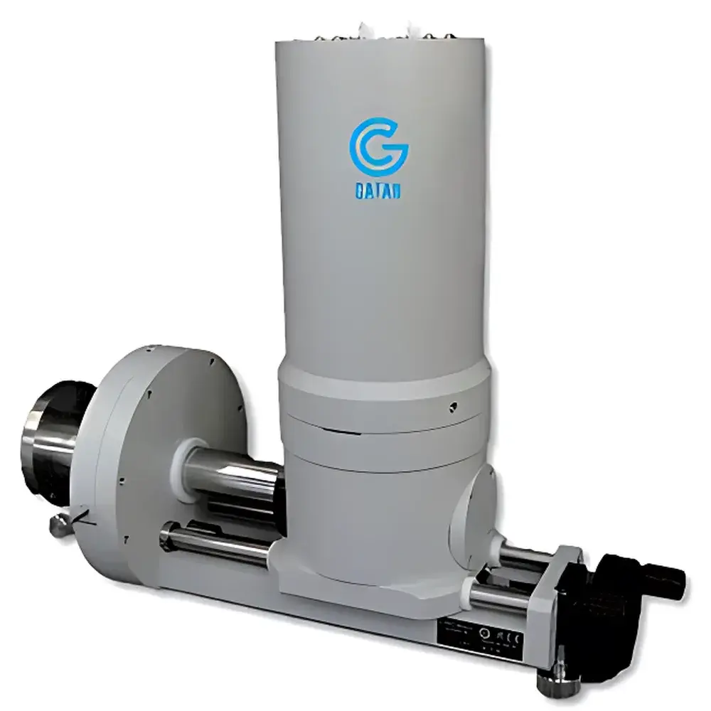
| Retraction Range | 150 mm |
| Integrated BSED Capability | Simultaneous Secondary Electron (SE), Backscattered Electron (BSE), and CL Acquisition |
| Mounting | Fixed Insertion Geometry with Lockable Retraction |
| Operating System | Windows 7 (32-/64-bit) |
Overview
The Gatan ChromaCL2 is a second-generation real-time color cathodoluminescence (CL) imaging system engineered for integration with scanning electron microscopes (SEM). It operates on the fundamental principle of cathodoluminescence—where incident high-energy electrons excite luminescent centers in insulating or semiconducting materials, resulting in photon emission across the visible and near-visible spectrum. Unlike monochromatic or sequential wavelength-scanning CL systems, the ChromaCL2 employs parallel spectral dispersion via a custom grating optical train coupled to an array photomultiplier tube (PMT), enabling simultaneous acquisition of full spectral information across ~400–800 nm within a single electron beam scan. This architecture eliminates time-dependent artifacts and ensures pixel-correlated color fidelity essential for quantitative mineralogical interpretation, trace-element zoning analysis, and defect mapping in geological and advanced materials specimens.
Key Features
- Single-Scan Real-Time Color Imaging: Full RGB-composite CL images generated in real time during beam rastering—no post-scan reconstruction or spectral stitching required.
- High-Efficiency Optical Collection: Parabolic mirror fabricated via precision diamond turning achieves large solid-angle collection (>0.15 sr), maximizing signal throughput while preserving spatial resolution down to sub-micron scales.
- Parallel Multichannel Detection: Grating-based spectral dispersion directs photons onto a linear array PMT, enabling concurrent detection across the entire visible range without mechanical filter wheels or tunable monochromators.
- Integrated Multi-Modal Signal Acquisition: Native synchronization with DigiScan II allows simultaneous acquisition of secondary electrons (SE), backscattered electrons (BSE), and CL signals—enabling direct correlation of structural, compositional, and optoelectronic contrast in one experiment.
- Phosphorescence Suppression Logic: Built-in pulse discrimination electronics distinguish prompt CL emission from delayed phosphorescent decay, critical for accurate imaging of halide-containing minerals (e.g., fluorite, apatite) and phosphor materials.
- Robust Mechanical Design: Motorized retraction mechanism with 150 mm travel range and fixed insertion geometry ensures repeatable positioning and minimizes alignment drift during long-term operation or multi-instrument sharing.
- Extended Violet Sensitivity: Optimized photocathode response and anti-reflection coatings extend usable spectral response into the near-UV (down to ~400 nm), supporting analysis of wide-bandgap semiconductors (e.g., AlN, GaN) and rare-earth-doped phosphors.
Sample Compatibility & Compliance
The ChromaCL2 is compatible with standard SEM specimen geometries up to 150 mm in diameter and accommodates both conductive and non-conductive geological, ceramic, semiconductor, and luminescent material samples. Conductive coating (e.g., carbon or Au/Pd sputtering) is recommended for insulating specimens to prevent charging during extended dwell times. The system complies with international standards governing analytical electron microscopy instrumentation, including ISO/IEC 17025 requirements for measurement traceability and ASTM E1508-22 for cathodoluminescence methodology in mineral identification. All acquired data—including raw pulse histograms, calibrated intensity maps, and metadata (beam kV, probe current, dwell time, stage coordinates)—are stored in DigitalMicrograph’s native DM3/DM4 format, supporting audit trails required under GLP and FDA 21 CFR Part 11 for regulated laboratories.
Software & Data Management
Data acquisition and visualization are fully integrated within Gatan’s DigitalMicrograph® platform, enhanced by the proprietary ChromaCL plugin. The software provides real-time RGB mixing algorithms that convert spectrally resolved photon counts into perceptually uniform color spaces (CIE XYZ or sRGB), with user-definable hue-saturation-luminance (HSL) mappings for targeted phase discrimination. Advanced features include automated mosaic stitching (Montage), spectral unmixing via linear combination fitting, region-of-interest (ROI) spectral extraction, and batch processing of time-series or tilt-series CL datasets. All image and spectrum files embed EXIF-style metadata—including instrument configuration, calibration timestamps, and operator ID—ensuring full experimental reproducibility and compliance with FAIR (Findable, Accessible, Interoperable, Reusable) data principles.
Applications
- Geosciences: Zoning analysis in zircon, monazite, and apatite for U–Pb geochronology; identification of fluid inclusion histories via healed microfractures; differentiation of detrital vs. authigenic quartz in shale reservoirs.
- Petroleum Geology: Pore-network characterization in carbonate and siliciclastic reservoir rocks; cementation sequence reconstruction using CL-intensity gradients and spectral shifts.
- Materials Science: Defect mapping in GaN-based LEDs and laser diodes; grain-boundary luminescence in polycrystalline perovskites; dopant distribution analysis in rare-earth-doped phosphors (e.g., YAG:Ce, SrAl₂O₄:Eu).
- Planetary Science: Comparative CL spectroscopy of extraterrestrial regolith simulants and meteoritic silicates to infer thermal and irradiation histories.
- Forensics & Cultural Heritage: Provenance determination of gemstones and archaeological ceramics through trace-element–sensitive CL signatures.
FAQ
What distinguishes ChromaCL2 from first-generation CL systems?
The ChromaCL2 replaces sequential filter-based or monochromator-scanned detection with true parallel spectral acquisition, eliminating temporal misregistration and enabling quantitative pixel-by-pixel spectral analysis within a single scan.
Is ChromaCL2 compatible with field-emission SEMs (FE-SEM)?
Yes—the system supports beam currents from 10 pA to >10 nA and is optimized for both thermionic and Schottky FE sources, with adjustable dwell time and line averaging to accommodate high-resolution mapping requirements.
Can CL spectra be exported for external analysis?
Yes—full spectral histograms (wavelength vs. counts) are exportable as ASCII or CSV files, retaining calibrated wavelength axes and absolute intensity scaling for use in third-party spectroscopic modeling tools.
Does the system support correlative light-electron microscopy (CLEM)?
While not a CLEM-dedicated platform, ChromaCL2-acquired CL maps can be registered to fluorescence or confocal images using DigitalMicrograph’s geometric transformation tools when fiducial markers or common structural features are present.
What maintenance is required for long-term stability?
The sealed optical path and solid-state electronics require no routine calibration; annual verification of PMT gain stability and grating alignment using NIST-traceable reference standards is recommended for GLP-compliant labs.


