PSI FluorCam-MultiSpectral Modular Plant Phenotyping Imaging System
| Brand | PSI (Czech Republic) |
|---|---|
| Origin | Czech Republic |
| Model | FluorCam-MultiSpectral Modular Plant Phenotyping Imaging System |
| Thermal Imager | Uncooled VOx microbolometer, 640×512 px, 7.5–13.5 µm, −25 to +150 °C, NETD ≤ 0.03 °C @ 30 °C |
| Fluorescence Imaging | UV/Blue/Red excitation, F440/F520/F690/F740 detection, 1360×1024 CCD, 20 fps |
| RGB Imaging | 2592×1944 sensor, 1–40× zoom, FOV 6.1×7.9 mm (40×) to 20.8×25.4 mm |
| NDVI/PAR Module | 630 nm & 740 nm LEDs |
| Max Sample Height | 50 cm |
| Compliance | CE-marked, ISO/IEC 17025 traceable calibration documentation available |
| Software | FluorCam v7.x with GLP-compliant audit trail, FDA 21 CFR Part 11 optional modules |
Overview
The PSI FluorCam-MultiSpectral Modular Plant Phenotyping Imaging System is a research-grade, multi-modal imaging platform engineered for quantitative, non-invasive phenotypic and physiological characterization of plants under controlled or semi-field conditions. It integrates three core optical modalities—multispectral chlorophyll fluorescence imaging, high-resolution thermal infrared imaging, and scientific-grade RGB macro-imaging—within a single, mechanically stable gantry-based architecture. The system operates on well-established biophysical principles: chlorophyll fluorescence emission (F440, F520, F690, F740) reports on photosynthetic electron transport efficiency, photoprotective dissipation, and secondary metabolite status; thermal infrared imaging (7.5–13.5 µm) captures stomatal conductance heterogeneity and water-use efficiency via leaf temperature gradients; and RGB imaging enables morphometric quantification (leaf area, perimeter, aspect ratio, color indices) at resolutions down to sub-millimeter scale. Designed for reproducible longitudinal studies, the system supports fully automated, time-series acquisition across multiple spectral domains, making it suitable for high-throughput screening in plant genetics, stress physiology, and functional genomics.
Key Features
- Modular hardware architecture: Independent, interlock-compatible units—Fluorescence Imaging Module, Thermal IR Imaging Module, RGB Macro-Imaging Module, and optional NDVI/PAR Absorption Module—allow configuration scalability without compromising optical alignment stability.
- UV-excited multispectral fluorescence imaging: Simultaneous or sequential acquisition of four emission bands (F440, F520, F690, F740) using calibrated narrowband filters and high-quantum-efficiency CCD sensors (1360×1024, 6.45 µm pixel size), enabling separation of epidermal/mesophyll-derived blue-green fluorescence (BGF) from chlorophyll-a-associated red/far-red fluorescence (Chl-F).
- Thermal imaging with metrological traceability: Uncooled VOx microbolometer (640×512 px) certified per EU standards for absolute radiometric accuracy; real-time temperature mapping with ±0.5 °C accuracy across −25 to +150 °C range; supported by atmospheric correction parameters (humidity, distance, emissivity) and IP65-rated environmental protection.
- Scientific RGB imaging: 2592×1944 CMOS sensor with 54 dB SNR and continuous 1–40× optical zoom; calibrated color reproduction; automated segmentation for leaf area, shape descriptors, greenness index (GI), and histogram-based color classification.
- Flexible excitation source library: Interchangeable LED panels including UV-A (365 nm), blue (450 nm), green (520 nm), red (630 nm), far-red (740 nm), and white-spectrum LEDs—enabling targeted interrogation of flavonoid accumulation, stomatal aperture dynamics, and PAR absorption kinetics.
- Automated protocol-driven operation: Predefined or user-defined experimental sequences (e.g., Fv/Fm, Kautsky induction, NPQ relaxation, light-response curves) executed unattended with programmable repetition intervals, timestamped metadata logging, and auto-save to hierarchical folder structures compliant with FAIR data principles.
Sample Compatibility & Compliance
The FluorCam-MultiSpectral accommodates intact plant specimens ranging from detached leaves and seedlings to whole rosettes and small canopy-level arrangements (up to 50 cm height). Its open gantry design permits integration with growth chambers, climate-controlled rooms, or greenhouse-mounted rails. All imaging modules comply with CE marking requirements and are supplied with factory calibration certificates traceable to national metrology institutes. Thermal imaging performance adheres to ISO 18434-1 for condition monitoring and ASTM E1933 for infrared thermography in biological applications. Fluorescence quantification protocols align with established plant physiology standards (e.g., OEC recommendations for chlorophyll fluorescence measurement). Optional software modules support 21 CFR Part 11 compliance—including electronic signatures, audit trails, and role-based access control—for laboratories operating under GLP or GMP frameworks.
Software & Data Management
FluorCam v7.x is a native Windows application built on a modular, extensible architecture. It provides four primary workflow environments: Live (real-time preview and manual ROI placement), Protocol (graphical editor for multi-step experiments), Pre-processing (background subtraction, flat-field correction, spectral unmixing), and Result (parameter extraction, statistical export, and visualization). Each image dataset is stored with embedded EXIF-style metadata: acquisition timestamp, excitation/emission wavelengths, exposure settings, ambient conditions, and operator ID. ROI analysis supports arbitrary geometric definitions (point, line, polygon, ellipse, freehand), with per-ROI statistics exported as CSV or Excel. Time-series data generate dynamic parameter plots (e.g., ΦPSII kinetics, Tleaf variance over time) and support batch processing across hundreds of images. Raw data files are saved in lossless TIFF format; processed results include annotated false-color overlays, histograms, scatter plots, and customizable PDF reports.
Applications
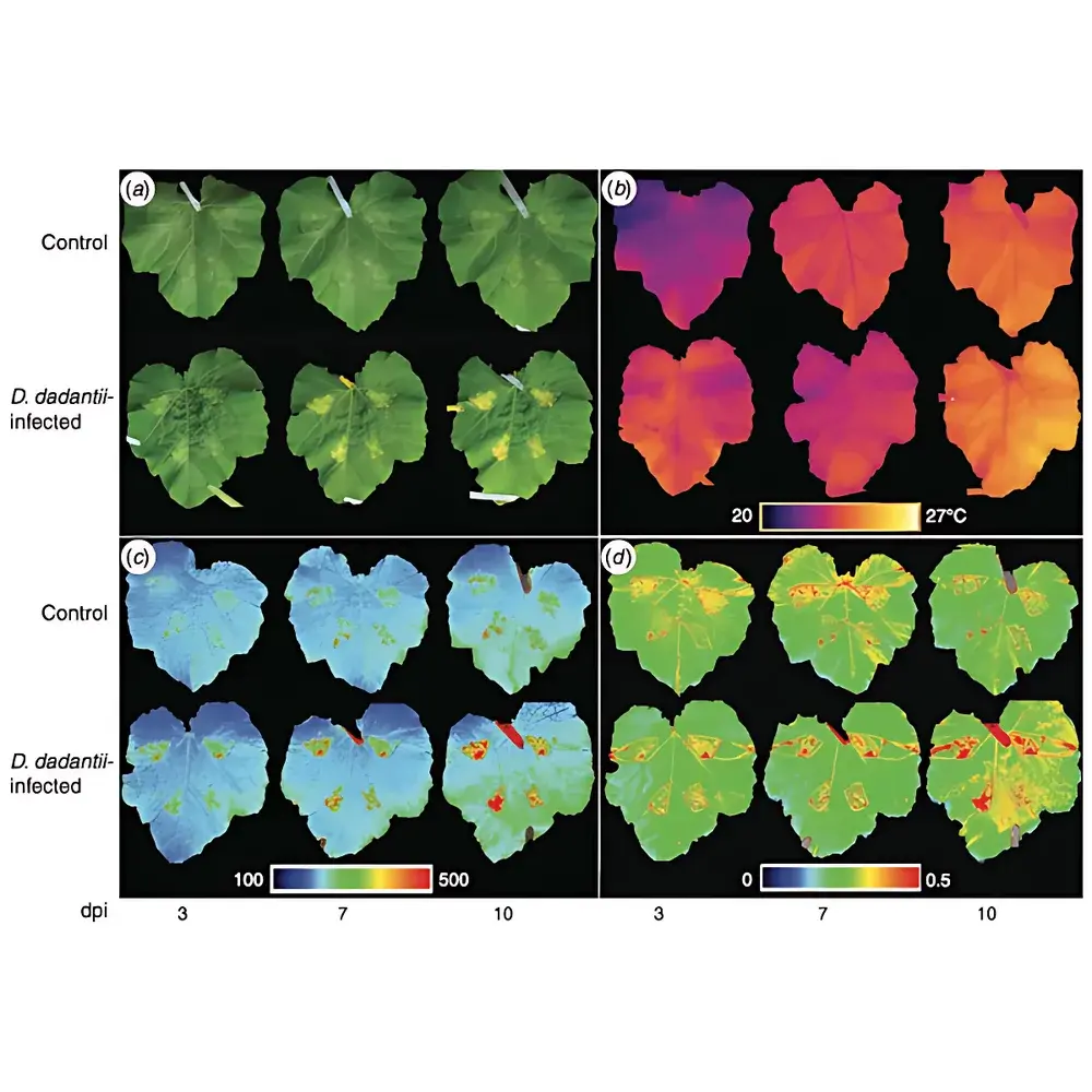
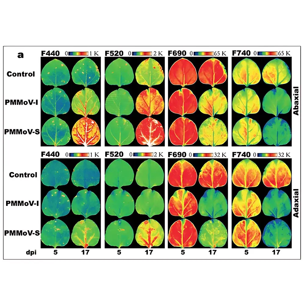
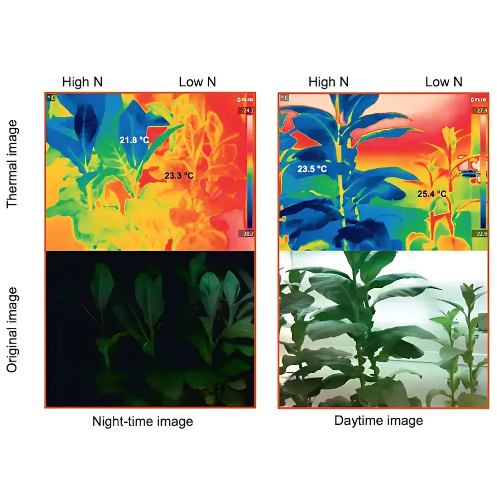
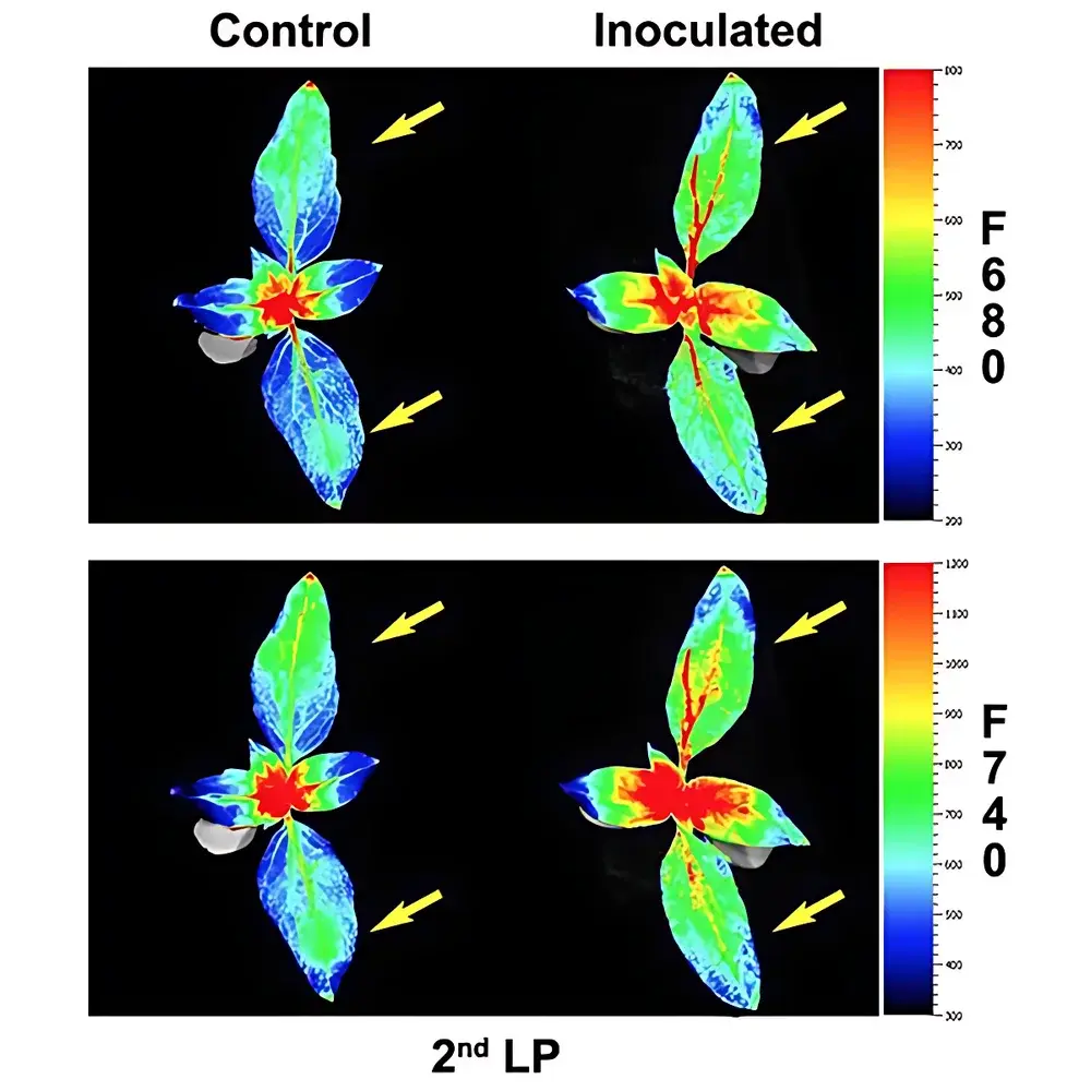
This system serves as a foundational tool across multiple domains in plant science. In crop breeding programs, it enables early-stage phenotyping of drought tolerance (via thermal heterogeneity), disease resistance (via multicolor fluorescence anomaly detection), and nitrogen use efficiency (via NDVI and Chl-F ratios). In functional genomics, it supports high-resolution mapping of QTLs associated with stomatal regulation, photochemical quenching, or anthocyanin biosynthesis. Stress physiology studies leverage its capacity to resolve spatiotemporal dynamics—for example, tracking Orobanche infection progression in sunflower through F690/F740 ratio shifts before visible symptoms appear. Published case studies demonstrate its utility in detecting Dickeya dadantii infection in zucchini via combined RGB/thermal/fluorescence pattern recognition, and in quantifying metabolic reprogramming during Pseudomonas syringae challenge in bean using spatially resolved F520/F690 maps. The platform also supports GFP/YFP reporter line validation and developmental trajectory analysis in model species such as Arabidopsis, tomato, and maize.
FAQ
What is the maximum sample height supported by the standard configuration?
The gantry accommodates plants up to 50 cm tall; custom-height extensions are available upon request.
Can the system be integrated into an existing growth chamber or greenhouse?
Yes—the modular design allows mounting on linear rails or fixed frames; electrical and data interfaces (USB 3.0, GigE) are compatible with industrial control systems.
Is spectral calibration performed per unit prior to shipment?
Each fluorescence module undergoes factory spectral calibration using NIST-traceable reference sources; thermal modules include individual pixel-wise radiometric calibration tables.
Does the software support machine learning-based classification workflows?
Raw image stacks and extracted feature tables (CSV/Excel) are fully compatible with Python (scikit-learn, TensorFlow), R, and MATLAB; example scripts for supervised classification of pathogen-infected vs. healthy tissue are provided in the user documentation.
How is data integrity ensured during long-term无人值守 experiments?
The software implements write-verification checksums, automatic folder timestamping, dual-storage options (local SSD + network backup), and configurable error logging with email alerts for acquisition failures.


