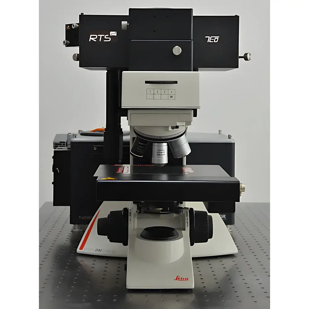
ZOLIX RTS2 Confocal Micro-Raman Spectroscopy System
| Brand | ZOLIX |
|---|---|
| Model | RTS2 |
| Instrument Type | Confocal Micro-Raman Spectrometer |
| Spectral Range | 300–1100 nm |
| Spectral Resolution | ≤1 cm⁻¹ |
| Spatial Resolution | Lateral ≤1 µm, Axial ≤2 µm |
| Minimum Wavenumber | 10 cm⁻¹ |
| Spectral Repeatability | ±0.1 cm⁻¹ |
| Laser Options | Integrated 532 nm DPSS (TEM₀₀, 100 mW), 638 nm & 785 nm diode lasers |
| Spectrometer | 328 mm focal length, four-grating turret (1800, 600, 150 l/mm + optional fourth grating) |
| Detector | 2000 × 256-pixel deep-cooled Raman-optimized CCD (QE >90% at peak), EMCCD/PMT upgradeable |
| Confocal Modes | Slit-CCD confocal and fiber-pinhole confocal (switchable) |
| Mapping Capability | Motorized XY stage with 2D Raman mapping (1 µm step resolution) |
| SNR | >20:1 (Si third-order peak) |
| Optional Accessories | Ultra-low wavenumber module (10 cm⁻¹), dark-field scattering spectroscopy, in-situ 2D photoluminescence (PL) and Raman co-mapping |
Overview
The ZOLIX RTS2 Confocal Micro-Raman Spectroscopy System is a research-grade, modular confocal Raman platform engineered for high-fidelity vibrational spectroscopic analysis at the microscale. Based on true confocal optical architecture—employing either slit-CCD or fiber-pinhole spatial filtering—the system delivers diffraction-limited lateral resolution (<1 µm) and sub-2 µm axial sectioning capability, enabling precise depth-resolved chemical characterization of heterogeneous samples. Its core optical design integrates a stabilized, compact Raman path with minimal beam walk-off, ensuring long-term spectral reproducibility (±0.1 cm⁻¹) and thermal drift resistance. The system operates across a broad spectral range (300–1100 nm), supporting multiple excitation wavelengths (532 nm, 638 nm, 785 nm) with TEM₀₀ beam quality and built-in laser alignment stability—eliminating routine realignment during multi-wavelength experiments. As a fully compatible add-on to unmodified research-grade upright microscopes, the RTS2 preserves all native microscope functionalities—including Köhler illumination, DIC, fluorescence, and transmitted light imaging—while adding quantitative Raman mapping, spectral correlation, and multimodal correlative analysis.
Key Features
- Modular confocal architecture with dual-path selection: high-sensitivity free-space coupling and high-axial-resolution fiber-pinhole confocal mode—switchable via software-controlled optics
- Integrated laser engine with factory-aligned 532 nm DPSS laser (100 mW, single longitudinal mode), plus selectable 638 nm and 785 nm diode lasers; optional single-mode fiber input for external laser integration
- 328 mm focal length spectrograph with motorized four-grating turret (1800, 600, 150 l/mm standard; fourth grating user-selectable), enabling optimized resolution/sensitivity trade-offs across UV–NIR
- Deep-cooled, back-illuminated scientific CCD detector (2000 × 256 pixels) with >90% peak quantum efficiency and thermoelectric cooling to –70°C; supports EMCCD and PMT detector upgrades for weak-signal or time-resolved applications
- Precision motorized XYZ stage with sub-micron step resolution, enabling automated 2D/3D Raman mapping, line scans, and hyperspectral data cube acquisition
- Patented TruRes spectral calibration technology and auto-focus algorithm ensure pixel-accurate spectral registration and consistent focus across large-area mapping
- Native compatibility with commercial upright microscopes (e.g., Nikon, Olympus, Zeiss); no optical modifications required—full retention of microscope functionality including phase contrast, polarized light, and epifluorescence
Sample Compatibility & Compliance
The RTS2 accommodates diverse solid, thin-film, powder, and biological specimens—including semiconductors, 2D materials (graphene, TMDCs), pharmaceutical polymorphs, geological inclusions, and fixed tissue sections—without destructive preparation. Its ultra-low wavenumber option (down to 10 cm⁻¹) enables detection of lattice modes, interlayer vibrations, and acoustic phonons critical in layered materials and soft matter. The system meets essential laboratory compliance prerequisites: spectral calibration traceability to NIST-traceable standards (e.g., silicon, neon lamps); wavelength accuracy certified per ISO 17025-accredited procedures; and full audit trail support for GLP/GMP environments when paired with compliant data management software. While not FDA 21 CFR Part 11–certified out-of-the-box, its raw data export (FITS, ASCII, HDF5) and metadata-rich acquisition logs facilitate integration into validated analytical workflows under regulated QA/QC frameworks.
Software & Data Management
Acquisition and analysis are performed via ZOLIX’s dedicated RamanStudio platform—a Windows-based application supporting real-time spectral preview, multi-channel synchronization (Raman/PL/reflectance), and batch processing of mapping datasets. The software implements ASTM E1840-compliant spectral preprocessing (cosmic ray removal, baseline correction via asymmetric least squares, intensity normalization), multivariate analysis (PCA, cluster analysis), and spectral library matching (user-expandable). All raw spectra retain embedded metadata (laser power, exposure time, grating, objective, stage coordinates, temperature log if interfaced), ensuring FAIR (Findable, Accessible, Interoperable, Reusable) data principles. Export formats include industry-standard .spc, .txt, .csv, and HDF5 for interoperability with MATLAB, Python (SciPy, scikit-learn), and commercial chemometrics packages. Audit trails, user access controls, and electronic signature options are available via optional enterprise licensing modules aligned with ISO/IEC 17025 documentation requirements.
Applications
The RTS2 serves as a primary tool in academic and industrial labs conducting advanced materials characterization, including strain and doping mapping in semiconductor heterostructures; polymorph identification and crystallinity assessment in pharmaceutical formulations; defect density quantification in CVD-grown graphene; stress/strain distribution analysis in microelectronic packaging; and label-free molecular histopathology of formalin-fixed paraffin-embedded (FFPE) tissue sections. Its dual confocal modes allow optimization for either maximum signal throughput (free-space) or stringent depth discrimination (fiber-pinhole)—critical for analyzing multilayer thin films, buried interfaces, or turbid biological matrices. The optional dark-field scattering module extends capability to plasmonic nanoparticle characterization, while in-situ 2D PL/Raman co-mapping enables simultaneous electronic and vibrational fingerprinting of optoelectronic devices under environmental control (temperature, gas atmosphere).
FAQ
What laser wavelengths are standard on the RTS2 system?
The system includes a factory-aligned 532 nm DPSS laser (100 mW, TEM₀₀) as standard. 638 nm and 785 nm diode lasers are available as configurable options. A single-mode fiber input port allows integration of external lasers (e.g., 488 nm, 660 nm, or tunable sources).
Can the RTS2 perform true 3D Raman tomography?
Yes—using the motorized Z-stage and fiber-pinhole confocal mode, the system acquires depth-resolved spectral stacks with axial resolution ≤2 µm (at 100× objective), enabling reconstruction of 3D chemical distribution volumes.
Is the system compatible with vacuum or controlled-atmosphere stages?
The optical interface is designed for standard microscope nosepiece mounting; any commercially available vacuum-compatible or environmental stage (e.g., Linkam, Instec) can be integrated provided mechanical and optical clearances are maintained.
Does the software support automated spectral library identification?
Yes—RamanStudio includes a customizable spectral library manager with correlation-based matching (Pearson, Euclidean, cosine similarity) and threshold-adjustable hit scoring, compliant with ASTM E1446 guidelines for spectral identification.
What is the typical acquisition time for a 100 × 100 µm Raman map at 1 µm step size?
At 1 s per spectrum (typical for moderate-signal samples using 532 nm excitation), a 100 × 100 point map requires ~2.8 hours; acquisition time scales linearly with integration time and inversely with laser power and detector sensitivity settings.