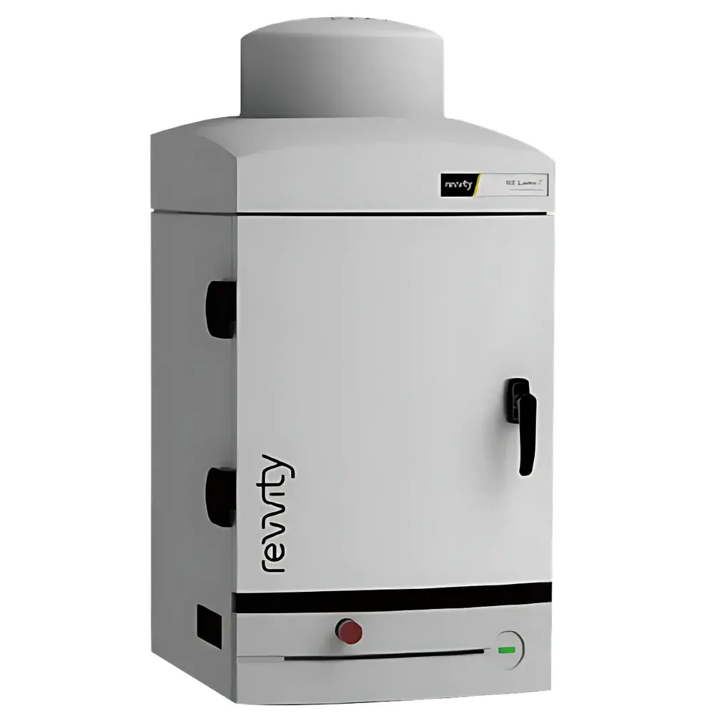
Revvity IVIS Lumina Series III Small Animal In Vivo Optical Imaging System
| Brand | Revvity |
|---|---|
| Origin | USA |
| Manufacturer | Revvity, Inc. |
| Product Type | In Vivo Optical Imaging System |
| Model | IVIS Lumina Series III |
| Imaging Modalities | Bioluminescence, Fluorescence (Multispectral), Cerenkov Luminescence |
| Animal Compatibility | Mouse (also supports rat, cell culture, microplates) |
| Field of View Options | 2.5 × 2.5 cm to 24 × 24 cm (motorized zoom lens with 5 preset FOVs) |
| Filter Wheel Capacity | 26 positions (standard) |
| CCD Sensor | Back-illuminated, deep-cooled scientific-grade CCD (–90 °C) |
| Lens Aperture | f/0.95 |
| Filter Transmission | ≥95% (excitation & emission) |
| Spectral Analysis | Integrated Continuous Pure Spectral (CPS) algorithm |
Overview
The Revvity IVIS Lumina Series III is a high-performance, multi-modal small animal in vivo optical imaging platform engineered for quantitative bioluminescence, fluorescence, and Cerenkov luminescence imaging. Built upon over two decades of IVIS platform development, the Series III integrates a back-illuminated, deep-cooled scientific CCD detector (operating at –90 °C), an ultra-fast f/0.95 lens assembly, and a motorized five-position field-of-view (FOV) system ranging from 2.5 × 2.5 cm to 24 × 24 cm. Its optical architecture is optimized for photon capture efficiency across visible and near-infrared (NIR) wavelengths (400–900 nm), enabling detection of low-intensity signals from single-cell bioluminescent tumors or deeply seated fluorescent probes. The system operates on fundamental principles of photon detection under controlled dark conditions—bioluminescence relies on enzymatic light generation (e.g., luciferase–luciferin reaction), fluorescence requires external excitation and wavelength-specific emission collection, and Cerenkov imaging captures visible photons emitted by high-energy beta emitters (e.g., 18F, 90Y) traveling faster than light in tissue. This tri-modal capability supports longitudinal, non-invasive monitoring of biological processes—including tumor progression, metastasis, gene expression, immune cell trafficking, and therapeutic response—in live murine models under standardized, GLP-compatible experimental conditions.
Key Features
- Deep-cooled back-illuminated CCD sensor operating at –90 °C to minimize dark current and read noise, ensuring high signal-to-noise ratio (SNR) even during long exposures.
- Motorized, five-step FOV system with calibrated magnification options (2.5 × 2.5 cm to 24 × 24 cm), supporting both high-resolution single-mouse imaging and parallel acquisition of up to five mice or two rats.
- 26-position automated filter wheel preloaded with high-transmission (>95%) excitation and emission filters, covering spectral bands from 430 nm to 840 nm.
- Integrated Continuous Pure Spectral (CPS) algorithm—the industry-recognized gold standard for multispectral unmixing—enabling robust autofluorescence subtraction, quantitative separation of multiple fluorophores (e.g., Cy5.5, IRDye 800CW, Alexa Fluor 750), and pixel-level spectral deconvolution.
- Dual-mode illumination: High-stability LED-based excitation source with uniform intensity distribution across the full spectral range; optional halogen lamp for broader excitation flexibility.
- Modular hardware design supporting ex vivo applications—including petri dishes, multi-well plates (6–384-well), tissue sections, and explanted organs—without reconfiguration.
Sample Compatibility & Compliance
The IVIS Lumina Series III is validated for use with immunocompromised and transgenic mouse models (e.g., NSG, SCID/beige, BALB/c), including orthotopic, subcutaneous, and metastatic tumor models. It accommodates standard anesthesia delivery systems (isoflurane-compatible stage with integrated gas ports) and temperature-controlled stages (37 °C ± 0.5 °C) to maintain physiological homeostasis during imaging sessions. All optical components undergo factory calibration traceable to NIST standards, ensuring inter-instrument data reproducibility—a critical requirement for multi-site preclinical studies and regulatory submissions. The system complies with ISO 13485 design controls for medical device-related research instrumentation and supports audit-ready electronic records when paired with optional 21 CFR Part 11-compliant software modules (e.g., Living Image® v4.7+ with audit trail, user access control, and electronic signature capabilities). It is routinely deployed in laboratories adhering to GLP, GCP, and AAALAC-accredited animal care protocols.
Software & Data Management
Acquisition and analysis are performed using Living Image® software (v4.7 or later), a validated, modular platform designed specifically for quantitative optical imaging. The software provides automated spectral unmixing via CPS, region-of-interest (ROI) quantification with background-subtracted radiance units (p/sec/cm2/sr), kinetic time-series analysis, and 3D surface topography mapping (when used with optional stereo camera module). Data export supports DICOM, TIFF, and HDF5 formats for integration into institutional LIMS or ELN systems. Raw image metadata—including exposure time, binning factor, FOV setting, filter pair, excitation power, and environmental parameters—is embedded in every saved file, satisfying FAIR (Findable, Accessible, Interoperable, Reusable) data principles. Optional network licensing enables centralized license management across core facilities, while role-based permissions ensure secure data handling in regulated environments.
Applications
- Oncology: Longitudinal tracking of luciferase-expressing xenografts (e.g., 4T1-luc2), evaluation of targeted therapeutics using NIRF-labeled antibodies (e.g., anti-HER2), and assessment of metabolic activity via 18F-FDG Cerenkov imaging.
- Immunology: In vivo visualization of adoptively transferred luciferase- or fluorescent-protein-labeled T cells, macrophages, or dendritic cells in inflammation or infection models.
- Gene Therapy & Reporter Assays: Quantitative monitoring of promoter activity, CRISPR/Cas9 editing efficiency, and viral vector biodistribution using dual-reporter constructs (e.g., firefly luciferase + mCherry).
- Drug Development: Pharmacokinetic/pharmacodynamic (PK/PD) profiling of fluorescently tagged drug candidates, including biodistribution, target engagement, and clearance kinetics.
- Microbiology: Real-time imaging of bacterial or fungal infections using lux- or GFP-expressing pathogens in sepsis or pneumonia models.
FAQ
What is the minimum detectable bioluminescent signal for a single 4T1-luc2 cell in vivo?
Under optimal conditions (anesthetized nude mouse, 1-minute exposure, f/0.95 lens, –90 °C CCD), the IVIS Lumina Series III reliably detects bioluminescent signal from a single viable 4T1-luc2 cell implanted subcutaneously—validated per manufacturer’s application note AN-IVIS-003.
Can the system perform true spectral imaging without filter-based acquisition?
No—the IVIS Lumina Series III performs multispectral imaging via sequential filter-based acquisition followed by CPS algorithmic unmixing. It does not incorporate dispersive optics (e.g., gratings or prisms) for continuous spectral sampling.
Is Cerenkov imaging quantitative across different radioisotopes?
Quantification is isotope-dependent and must be calibrated using phantom studies. The system reports Cerenkov signal in radiance units, but absolute activity conversion requires co-registered PET/SPECT validation or dose-calibrated phantoms for each radionuclide (e.g., 18F vs. 89Zr).
Does the system support automated batch processing of multi-well plate data?
Yes—Living Image® includes dedicated microplate analysis workflows with well-defined ROI templates, background correction algorithms, and export to CSV/Excel for HTS-compatible downstream analysis.
How often does the system require optical recalibration?
Factory calibration is permanent and traceable; however, users are advised to perform quarterly performance verification using the included NIST-traceable reference standards (bioluminescent and fluorescent) to ensure ongoing compliance with internal QC protocols.


