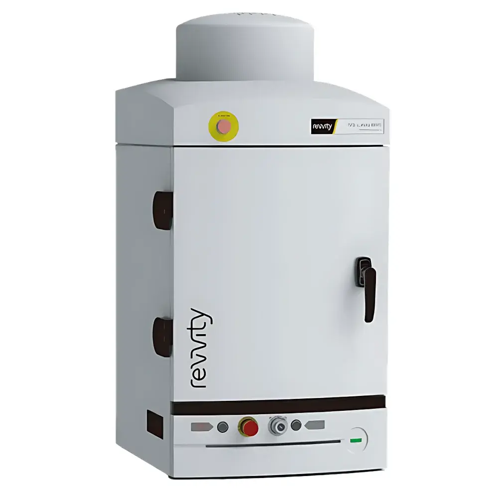
Revvity IVIS Lumina XRMS Small Animal In Vivo Optical Imaging System
| Brand | Revvity |
|---|---|
| Origin | USA |
| Manufacturer Type | Original Equipment Manufacturer (OEM) |
| Product Category | Imported Instrument |
| Model | Lumina XRMS |
| Instrument Type | Optical Imaging System |
| Animal Models | Mouse, Rat |
| Imaging Modalities | Bioluminescence, Fluorescence, Cherenkov, X-ray |
Overview
The Revvity IVIS® Lumina XRMS is a high-performance, multi-modal preclinical in vivo imaging platform engineered for quantitative, non-invasive longitudinal monitoring of biological processes in small animal models. Built upon proven IVIS optical imaging architecture, the Lumina XRMS integrates high-sensitivity bioluminescence detection, spectral unmixing-enabled fluorescence imaging, and low-dose, high-resolution X-ray radiography within a single instrument enclosure. Its core measurement principle combines photon detection via back-illuminated, thermoelectrically cooled CCD technology (optimized for 300–950 nm spectral range) with dual-energy X-ray acquisition using a flat-panel detector—enabling precise anatomical registration of optical signals. Designed specifically for mouse and rat studies, the system supports both 2D planar quantification and co-registered structural-functional analysis without requiring repositioning or inter-system transfer.
Key Features
- Multi-modal integration: Simultaneous or sequential acquisition of bioluminescence, fluorescence (with real-time spectral unmixing across 4–8 emission bands), Cherenkov luminescence, and X-ray radiography
- X-ray subsystem: 40–90 kVp adjustable tube voltage, <10 µm effective pixel size, dose-controlled exposure (<0.5 rad per image), and automatic bone/soft-tissue contrast optimization
- Optical sensitivity: Detection limit ≤100 photons/sec/cm²/sr for bioluminescence; fluorescence sensitivity down to 1–5 pM fluorophore in vivo depending on wavelength and depth
- Automated stage: Motorized, precision-aligned animal positioning with temperature-controlled (28–40 °C) heated platform and gas anesthesia integration (isoflurane compatible)
- Robust optical path: Vacuum-sealed, light-tight chamber with vibration-damped optical bench; calibrated reference standard for absolute radiance quantification
- Regulatory-ready design: Compliant with IEC 61000-6-3 (EMC), UL 61010-1 (safety), and supports 21 CFR Part 11-compliant audit trails when used with Living Image® Software v4.7+
Sample Compatibility & Compliance
The IVIS Lumina XRMS accommodates live mice (up to 50 g) and rats (up to 500 g) under controlled physiological conditions. It supports standard laboratory strains (e.g., C57BL/6, BALB/c, nude, NSG) and transgenic reporter models expressing luciferase (Firefly, Renilla, NanoLuc), fluorescent proteins (GFP, mCherry, iRFP720), or radiolabeled tracers (e.g., 89Zr, 64Cu). All optical and X-ray protocols are optimized to meet ALARA (As Low As Reasonably Achievable) radiation safety principles. The system conforms to ISO 13485 quality management standards for medical device manufacturing and supports GLP-compliant study execution per OECD Test Guidelines 407, 422, and 452 when paired with validated SOPs and instrument qualification documentation.
Software & Data Management
Imaging acquisition, processing, and analysis are performed using Living Image® Software—a validated, FDA 21 CFR Part 11-compliant platform featuring role-based user access control, electronic signatures, and full audit trail functionality. The software provides automated background subtraction, spectral unmixing algorithms (including linear unmixing and constrained least-squares fitting), region-of-interest (ROI) quantification with normalization to area or body weight, and 3D surface reconstruction from X-ray-guided optical signal localization. Raw data are stored in vendor-neutral formats (e.g., .ivis, DICOM for X-ray), enabling interoperability with third-party analysis tools such as MATLAB, Python (via PyIVIS), and commercial image analysis suites. Data export supports CSV, TIFF, and PDF reporting templates aligned with journal submission requirements (e.g., Nature Methods, PLOS ONE).
Applications
The IVIS Lumina XRMS serves as a foundational tool across oncology (tumor growth kinetics, metastasis tracking, therapy response assessment), immunology (cell trafficking, inflammation dynamics), infectious disease (pathogen burden, host response), neuroscience (neuroinflammation, gene therapy biodistribution), and drug development (PK/PD modeling, target engagement verification). Its ability to co-register functional optical signals with anatomical X-ray landmarks enables accurate spatial mapping of signal origin—critical for distinguishing superficial autofluorescence from deep-tissue reporter expression. The platform has been cited in over 15,000 peer-reviewed publications and supports regulatory submissions including IND-enabling toxicology studies and mechanistic pharmacology packages accepted by FDA and EMA.
FAQ
What is the minimum detectable bioluminescent flux for the Lumina XRMS?
The system achieves sub-picoWatt sensitivity—typically detecting ≥100 photons/sec/cm²/sr under standard acquisition settings (1–5 min exposure, f/1 lens, −90 °C sensor cooling).
Can the X-ray module be used for longitudinal bone density analysis?
Yes—the X-ray subsystem supports serial densitometry via calibrated Hounsfield unit (HU) referencing and ROI-based grayscale intensity tracking across timepoints.
Is spectral unmixing supported for all fluorescent probes?
Unmixing is validated for common near-infrared dyes (e.g., Cy5.5, IRDye800CW) and fluorescent proteins emitting between 480–840 nm; probe-specific spectral libraries must be pre-acquired or imported.
Does the system support GMP-compliant workflows?
When deployed with Living Image v4.7+ and documented IQ/OQ/PQ protocols, the Lumina XRMS meets key elements of GMP Annex 11 for computerized systems used in nonclinical safety studies.
How is animal temperature maintained during extended imaging sessions?
A closed-loop feedback system regulates platform temperature between 28–40 °C using PID-controlled heating elements and real-time rectal or surface thermistor monitoring.


