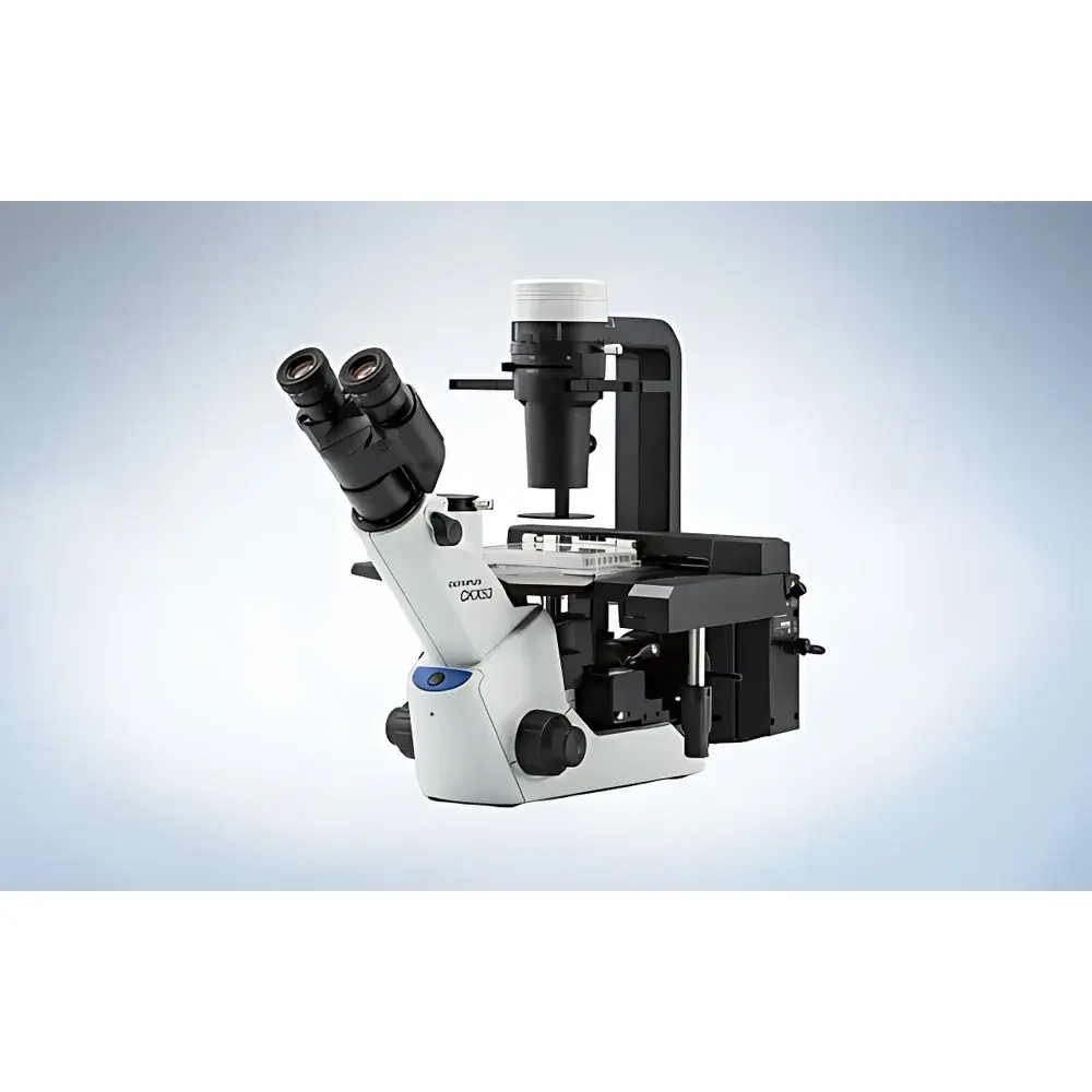
Olympus CKX53 Inverted Microscope
| Brand | Olympus |
|---|---|
| Origin | Beijing, China |
| Model | CKX53 |
| Instrument Type | Inverted Microscope |
| Optical System | UIS2 |
| Illumination | Long-life LED (white light), Optional 100W Mercury Lamp (U-LH100HG) for Fluorescence |
| Phase Contrast | Integrated iPC (intelligent Phase Contrast) System with Universal Annulus Plate for 10×, 20×, and 40× Objectives |
| IVC (Inverse Contrast) Technology | Enabled with PLCN10× or CACHN10×IPC Objective |
| Field Number | FN 22 |
| Weight | ~7 kg |
| Stage Compatibility | Standard CKX-MVR Manual Stage |
| Fluorescence Mirror Units | Interchangeable, Compatible with IX3/BX3 Series Filter Sets |
| UV-Resistant Coating | Yes |
| Sterility-Friendly Design | Bottom-mounted shutter, compact footprint, cleanbench-compatible |
Overview
The Olympus CKX53 is an engineered inverted microscope designed specifically for routine cell culture workflows in academic, pharmaceutical, and biomanufacturing laboratories. Built upon the proven UIS2 optical platform, it delivers high-fidelity, high-contrast imaging across multiple contrast modalities—including brightfield, phase contrast (iPC), inverse contrast (IVC), and fluorescence—without mechanical realignment or objective swapping. Its optical architecture employs Köhler illumination principles with a long-life white LED light source (rated >10,000 hours), ensuring stable color temperature (~5,800 K), uniform illumination across the full FN22 field of view, and minimal thermal load on live specimens. The iPC system integrates a universal annulus plate compatible with 10×, 20×, and 40× phase objectives, enabling rapid, reproducible phase contrast acquisition without manual ring alignment. Complementing this, the proprietary IVC technique—based on asymmetric illumination and differential interference principles—generates artifact-free, halo-free pseudo-3D visualization of transparent, unstained cells, as validated in peer-reviewed optical literature (Optics Letters, 2015). This makes the CKX53 particularly suited for longitudinal monitoring of adherent mammalian cultures, stem cell differentiation assays, and primary neuron morphology studies where label-free, non-invasive observation is critical.
Key Features
- Compact, low-profile chassis (≈7 kg) optimized for laminar flow hoods and biosafety cabinets—features bottom-mounted shutter and UV-resistant surface coating for compatibility with routine UV sterilization protocols.
- Integrated iPC system with pre-centered universal phase annulus for seamless switching between 10×, 20×, and 40× phase contrast objectives—eliminates iterative centering and improves inter-user reproducibility.
- IVC (Inverse Contrast) technology enabled via dedicated 10× objectives (PLCN10×, CACHN10×IPC), delivering enhanced depth perception and edge definition without directional shadows or phase halos—ideal for visualizing confluent monolayers and spheroid peripheries.
- Standardized C-mount camera port (23.2 mm) supporting OEM and third-party digital cameras; enables simultaneous acquisition in brightfield, phase, IVC, and fluorescence modes under ambient lab lighting.
- CKX-MVR manual stage with 70 mm lateral translation range and precision 90°-indexed movement for 96-well plates—enables rapid, repeatable well-to-well navigation without stage recalibration.
- Fluorescence-ready configuration with U-LH100HG 100 W mercury lamp and interchangeable mirror units compatible with IX3/BX3 filter sets—supports DAPI, FITC, TRITC, and mCherry excitation/emission profiles with high signal-to-noise ratio even at low-expression levels.
Sample Compatibility & Compliance
The CKX53 accommodates a broad spectrum of cell culture formats without adapter modification: standard 35 mm Petri dishes (up to three concurrently), 6–96-well microplates, T-25 to T-225 flasks, and multilayer vessels up to 190 mm in height (achieved by removing the condenser assembly). Its elevated specimen height capacity (19 mm working distance with UPLFLN4×IPC objective) permits imaging of lower layers in triple-layer flasks—a capability essential for high-density bioproduction monitoring. All optical components comply with ISO 10934-1 (microscope nomenclature) and JIS B 7151 (optical performance testing). While not certified for GMP production environments, its design supports GLP-aligned documentation practices: consistent illumination output, traceable focus mechanics, and stable stage positioning enable audit-ready image capture logs when paired with Olympus cellSens software.
Software & Data Management
The CKX53 interfaces natively with Olympus cellSens Entry and cellSens Standard software (Windows OS), providing calibrated measurement tools (area, length, intensity profiling), time-lapse scheduling, multi-channel fluorescence overlay, and metadata tagging (objective, contrast mode, exposure time, date/time stamp). Export formats include TIFF (16-bit), JPEG2000, and AVI—fully compatible with downstream analysis platforms such as ImageJ/Fiji, MATLAB, and commercial AI-based cell segmentation tools. When deployed in regulated environments, cellSens supports user access control, electronic signatures, and 21 CFR Part 11-compliant audit trails (requires licensed cellSens Dimension module). Raw image files retain embedded EXIF-style metadata—including illumination intensity setting, iPC/IVC activation status, and objective identification—facilitating retrospective protocol validation.
Applications
- Cell Culture Quality Control: Daily confluence assessment, morphology screening, and contamination detection in CHO, HEK293, iPSC, and primary fibroblast lines using label-free iPC and IVC.
- Transfection & Passaging Support: Real-time guidance during trypsinization, seeding density optimization, and clonal isolation—enhanced by wide-field FN22 optics and ergonomic stage controls.
- Fluorescent Reporter Monitoring: Tracking GFP/RFP expression kinetics, calcium flux (e.g., Fluo-4), or mitochondrial membrane potential (TMRM) in live cultures under controlled ambient lighting.
- Microplate-Based Assays: High-throughput viability scoring (e.g., Calcein-AM/EthD-1), spheroid size quantification, and neurite outgrowth analysis in 96-well formats.
- Educational & Core Facility Use: Robust, low-maintenance platform for undergraduate labs and shared imaging facilities—minimizes training overhead while maintaining quantitative comparability across users.
FAQ
Is the CKX53 suitable for long-term live-cell time-lapse imaging?
Yes—when paired with an environmental chamber (e.g., Tokai Hit INU series) and appropriate CO2/temperature control, the CKX53’s LED illumination generates negligible heat drift, supporting hour-scale acquisitions with sub-micron focus stability.
Can I use third-party fluorescence filter cubes with the CKX53?
Yes—the mirror unit slide accepts standard 25 mm diameter cubes conforming to Olympus IX3/BX3 mechanical dimensions and optical path lengths; verify transmission spectra compatibility prior to integration.
Does the CKX53 support motorized Z-focus or automated stage functions?
No—the base CKX53 is manually operated; however, Olympus offers the CKX53-MVRS variant with motorized XY stage and Z-drive for automated scanning workflows (sold separately).
What is the maximum vessel height supported without condenser removal?
With condenser installed, the working height clearance is ≈75 mm; removing the condenser extends this to 190 mm—sufficient for Corning CellSTACK® chambers and similar multilayer systems.
Is service and calibration documentation available for regulatory submissions?
Olympus provides factory calibration certificates (illuminance uniformity, magnification accuracy, stage linearity) and optional IQ/OQ protocols aligned with ISO/IEC 17025 standards—contact authorized service partners for site-specific qualification support.
Related Products


