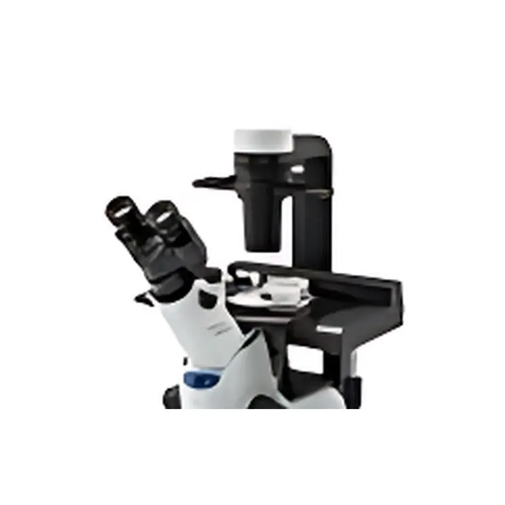
OLYMPUS CKX41 Inverted Biological Microscope
| Brand | OLYMPUS |
|---|---|
| Origin | Japan |
| Model | CKX41, CKX53 |
| Microscope Type | Inverted Microscope |
| Eyepiece Configuration | Trinocular |
| Optical System | UIS2 Infinity-Corrected Optics |
| Focusing Mechanism | Objective Nosepiece-Driven Coaxial Coarse/Fine Focus (Fixed Stage) |
| Total Focus Travel | +7 mm / –2 mm from 1 mm above stage surface |
| Coarse Focus Travel per Revolution | 39.6 mm |
| Fine Focus Travel per Revolution | 0.2 mm |
| Fine Focus Resolution | 2 µm |
| Objective Turret | 4-Position |
| Stage | Mechanical XY Stage (X = 120 mm, Y = 78 mm) with Removable Circular Insert (Ø25 mm) and Petri Dish Support (Ø35 mm) |
| Condenser | Removable Long Working Distance Condenser (NA 0.3, WD 72 mm) |
| Phase Contrast | Pre-centered and Centerable Phase Contrast Slides (PHC-Compatible Objectives: CPLN10×PH, CPLFLN10×PH, LCACHN20×PH) |
| Fluorescence Illumination | Vertical Fluorescence Unit with 50 W Mercury Lamp, Three-Position Filter Slider (B, G, Transmitted Light or U), Optional U-Filter Block |
| Eyepiece Options | Widefield WH10× (F.N. 22), WHB10× (F.N. 20), PWH10×, 35WH10× |
| Tube Options | U-CTR30-2 Trinocular Tube (30° inclination, 50–75 mm IPD, beam splitter 50/50 or 20/80), CKX-TBI Adjustable-Incline Binocular Tube (30°–60°), UTBI-3 (5°–35°) |
| Power Supply | Dual Voltage Auto-Switching (100–240 V AC, 50/60 Hz), Continuously Variable Intensity Control |
Overview
The OLYMPUS CKX41 Inverted Biological Microscope is engineered for routine and advanced live-cell observation in cell culture laboratories, quality control environments, and academic teaching facilities. Its inverted optical architecture positions the objective lenses beneath the specimen stage—enabling direct, high-stability imaging of cells growing in standard tissue culture vessels (e.g., flasks, Petri dishes, multi-well plates) without disturbing sterility or requiring sample transfer. Central to its performance is the UIS2 (Universal Intermediate System 2) infinity-corrected optical platform, which delivers exceptional flat-field correction, high contrast, and consistent resolution across the entire field of view (up to F.N. 22). The system supports seamless integration of phase contrast, optional fluorescence, and differential interference contrast (DIC) techniques—without compromising parfocality or introducing magnification drift when accessories are inserted into the parallel light path between objective and tube lens.
Key Features
- UIS2 Infinity-Corrected Optics: Ensures superior chromatic and spherical aberration correction, extended depth of field, and uniform illumination across wide-field eyepieces—critical for quantitative assessment of confluent monolayers and heterogeneous co-cultures.
- Pre-Centered Phase Contrast Slides: Eliminate manual centration for 4×, 10×, 20×, and 40× objectives; single-phase ring alignment enables rapid switching between magnifications with no optical recalibration required.
- PHC (Phase Contrast High Contrast) Objectives: Specifically designed to minimize edge distortion caused by meniscus effects at vessel boundaries—delivering sharp, artifact-free images of cells near dish rims and within microfluidic chambers.
- Adjustable-Incline Binocular & Trinocular Tubes: CKX-TBI and U-CTR30-2 tubes support ergonomic operation under biosafety cabinets (30°–60° tilt range, 48–75 mm IPD adjustment), while trinocular configurations provide dedicated port access for digital cameras compliant with C-mount or F-mount standards.
- Modular Fluorescence Capability: Vertical illuminator accepts standardized UIS2 filter cubes (B, G, U excitation); integrated mercury lamp housing ensures stable UV–visible output and thermal management for reproducible signal intensity over extended acquisition periods.
- Compact, Low-Profile Chassis: Minimal footprint (W × D × H ≈ 320 × 420 × 410 mm) facilitates integration with environmental chambers, micromanipulators, and laminar flow hoods—reducing workflow interruption during time-lapse experiments.
Sample Compatibility & Compliance
The CKX41 accommodates a broad range of biological specimens housed in standard labware: T-25/T-75 flasks, 35 mm and 60 mm Petri dishes, 24-/96-well plates, hemocytometers, and glass-bottom dishes. Its long-working-distance condenser (72 mm) and PHC objectives ensure optimal clearance for thick vessels and temperature-controlled stages. All optical components comply with ISO 10934-1 (Microscopes — Nomenclature of parts) and JIS B 7151 (Japanese Industrial Standard for Microscope Performance Testing). When configured with audit-trail-enabled digital imaging software (e.g., OLYMPUS cellSens), the system supports GLP/GMP-aligned documentation workflows—including user authentication, timestamped metadata embedding, and electronic signature capability per FDA 21 CFR Part 11 requirements.
Software & Data Management
The CKX41 serves as a hardware foundation for scalable digital microscopy workstations. Its trinocular port supports USB 3.0 or HDMI-compatible CMOS sensors (e.g., OLYMPUS UC90, DP27), enabling real-time image capture, measurement (area, length, particle count), and annotation. Integration with OLYMPUS cellSens Dimension software provides Z-stack acquisition, multi-channel fluorescence registration, and batch-processing pipelines compatible with TIFF, JPEG2000, and OME-TIFF formats. Exported datasets retain EXIF-compliant metadata (objective ID, magnification, exposure time, gain setting), facilitating traceability in regulatory submissions and peer-reviewed publication.
Applications
- Routine monitoring of adherent mammalian cell lines (HEK293, HeLa, CHO) during subculturing and passaging
- Assessment of primary neuron morphology, dendritic spine density, and glial activation in co-culture models
- Quality assurance of stem cell differentiation protocols using morphological scoring criteria (e.g., embryoid body compactness, ectoderm/mesoderm marker distribution)
- Time-lapse imaging of wound-healing assays, mitotic progression, and apoptosis kinetics under controlled CO2/temperature conditions
- Fluorescent validation of transfection efficiency (GFP/RFP reporters), calcium flux (Fluo-4), or mitochondrial membrane potential (JC-1)
- Educational demonstration of phase contrast principles, motility analysis, and basic histopathology using unstained tissue explants
FAQ
Is the CKX41 compatible with third-party digital cameras?
Yes—via standard C-mount or F-mount adapters; mechanical and electrical synchronization requires verification of shutter trigger latency and sensor cooling specifications.
Can the CKX41 perform DIC or Hoffman modulation contrast?
No—DIC requires specialized prisms and polarizers not supported by the CKX series; however, RC (Rheinberg Contrast) and floating-phase contrast modes are available through optional UIS2-compatible accessories.
What is the maximum working distance achievable with the standard condenser?
72 mm—sufficient for most commercial incubator-style stages and heated chamber inserts.
Does the CKX41 meet CE or UL safety certification requirements?
Yes—the instrument carries CE marking per EU Directive 2014/30/EU (EMC) and 2014/35/EU (LVD), and complies with UL 61010-1 for laboratory electrical equipment.
Are replacement parts and service manuals available internationally?
OLYMPUS maintains an authorized global service network; original spare parts (e.g., lamp housings, phase sliders, objective lenses) are distributed through certified distributors with documented calibration traceability to NIST standards.
Related Products


