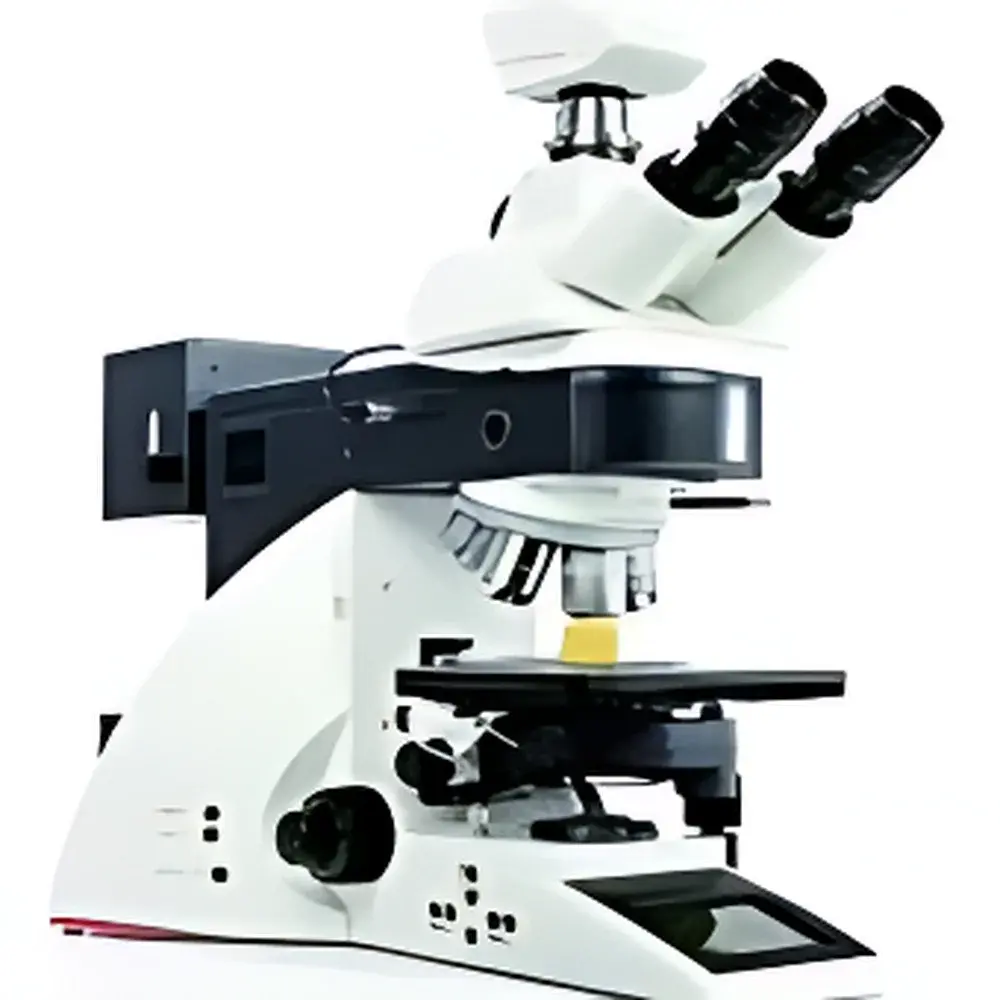
Leica DM4000M Semi-Automatic Metallurgical Microscope
| Brand | Leica |
|---|---|
| Origin | Germany |
| Model | DM4000M |
| Imaging Interface | FireWire (IEEE 1394) |
| CCD Resolution Options | 1.3 MP to 12 MP |
| Color Depth | 14-bit |
| Illumination Modes | Reflected & Transmitted Light |
| Automated Components | Motorized Filter Turret (4-position), Encoded Revolving Nosepiece (6-position), Motorized Field & Aperture Diaphragms, Motorized Condenser, Auto-Intensity Management (Reflected & Transmitted), Precise CCT Stabilization |
| Observation Modes | Brightfield, Darkfield, Polarization, DIC, Phase Contrast (transmitted), Fluorescence (reflected) |
| Display | Integrated LCD Status Panel |
| Software Control | Leica Application Suite (LAS) X-compatible, Free Download Available |
| Compliance | Designed for ISO/IEC 17025-compliant labs, supports GLP/GMP audit trails via optional LAS X modules |
Overview
The Leica DM4000M Semi-Automatic Metallurgical Microscope is an engineered solution for precision microstructural analysis of opaque, reflective specimens—primarily metals, alloys, ceramics, composites, electronic packaging, and powder metallurgy samples. Unlike conventional optical microscopes, the DM4000M employs a fully integrated upright optical path optimized for epi-illumination (reflected light), with optional dual-path capability supporting both reflected and transmitted illumination. Its core design principle centers on measurement repeatability and operator-independent condition replication: every imaging parameter—including illumination intensity, color temperature, diaphragm settings, objective position, and contrast mode—is digitally encoded, motorized, and recallable via hardware or software. This architecture ensures that identical acquisition conditions can be restored with ≤0.5% photometric deviation across sessions—critical for comparative analysis, quality control documentation, and regulatory submissions.
Key Features
- Motorized 6-position encoded nosepiece with automatic objective identification and magnification reporting on the integrated LCD panel
- 4-position motorized reflector turret enabling rapid, repeatable switching between brightfield, darkfield, polarization, fluorescence, and differential interference contrast (DIC)
- Auto-intensity management system dynamically adjusts LED or halogen illumination output in real time to maintain consistent irradiance across magnifications and objectives
- Constant Correlated Color Temperature (CCT) control (6500 K ±50 K) ensures photometric stability for quantitative colorimetric analysis and archival imaging
- Motorized field and aperture diaphragms synchronized with objective selection to optimize depth of field and resolution per magnification
- Motorized condenser with automated alignment for transmitted-light modes (phase contrast, polarized light, brightfield/darkfield)
- FireWire (IEEE 1394a) interface supporting real-time 14-bit image streaming at full frame rate—compatible with Windows and macOS platforms without proprietary drivers
- Modular platform supporting upgrade paths to automated stage control, focus mapping, and extended-depth-of-field (EDF) reconstruction
Sample Compatibility & Compliance
The DM4000M accommodates standard metallurgical specimen formats: 25 mm, 30 mm, and 50 mm diameter mounts; polished cross-sections up to 75 × 50 mm; and unmounted bulk samples via adjustable mechanical stage clamps. It complies with ISO 80000-7 (optical quantities), ISO 10934-1 (microscopy terminology), and ASTM E3 (Standard Guide for Preparation of Metallographic Specimens). When operated with Leica LAS X software and enabled audit trail modules, the system meets data integrity requirements under FDA 21 CFR Part 11 for electronic records and signatures—supporting user authentication, session logging, parameter change tracking, and immutable image metadata embedding. Its optical calibration traceability aligns with ISO/IEC 17025 accreditation criteria for testing laboratories performing microstructural evaluation.
Software & Data Management
Leica Application Suite (LAS) X provides native integration with the DM4000M’s motorized functions, enabling script-based acquisition protocols, multi-channel fluorescence registration, and batch image annotation compliant with MIAME/MINSEQE metadata standards. All captured images retain embedded EXIF-style metadata: objective ID, illumination mode, diaphragm values, exposure time, white balance coefficients, and system serial number. LAS X supports DIC quantitative phase gradient calculation, grain size analysis per ASTM E112, inclusion rating per ASTM E45, and hardness impression measurement per ISO 6507. Raw image data is stored in TIFF or Leica’s lossless LIF format, preserving 14-bit dynamic range for post-acquisition linear scaling and histogram equalization.
Applications
The DM4000M serves as a primary tool in failure analysis laboratories evaluating fatigue cracks, intergranular corrosion, weld microstructures, and coating delamination. In R&D environments, it supports phase identification in multiphase alloys (e.g., austenite/ferrite ratio quantification), ceramic grain boundary characterization, and semiconductor package defect inspection (voids, wire bond integrity, mold compound cracking). Quality assurance teams deploy it for routine verification of heat treatment homogeneity, surface finish assessment (Ra/Rz correlation), and particulate contamination analysis in cleanroom-assembled components. Its fluorescence capability enables oxide layer thickness estimation via interference color mapping, while polarization modes facilitate stress-induced birefringence detection in tempered glass or polymer films.
FAQ
Does the DM4000M support automated focus mapping for rough or tilted samples?
Yes—when paired with the optional motorized Z-drive and LAS X Focus Map module, the system acquires z-stacks and generates topographic surface models with sub-micron axial resolution.
Can illumination intensity be calibrated traceably to NIST standards?
Leica offers factory calibration certificates for the integrated photometric sensor; users may perform in-house verification using calibrated silicon photodiodes per ISO/IEC 17025 procedures.
Is DIC contrast quantifiable for strain analysis?
LAS X includes gradient vector field computation from DIC image pairs, enabling qualitative strain localization; quantitative strain mapping requires external calibration with known grating standards.
How is objective-specific optical correction maintained during automated mode switching?
Each encoded objective stores its numerical aperture, working distance, and tube lens compensation data; the system auto-adjusts condenser height and diaphragm geometry accordingly.
What cybersecurity measures are implemented for network-connected operation?
LAS X supports role-based access control, TLS 1.2 encrypted remote sessions, and optional integration with enterprise Active Directory—no default credentials or exposed telnet/FTP services.


