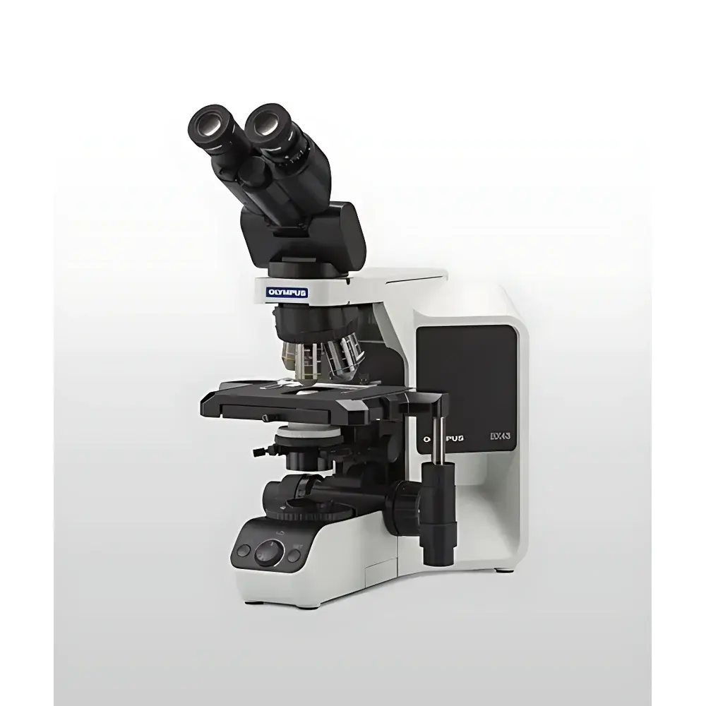
Olympus BX43 Upright Fluorescence Microscope
| Brand | Olympus |
|---|---|
| Origin | Japan |
| Model | BX43 |
| Type | Upright Biological Microscope |
| Illumination | LED Transmitted Köhler Illuminator (White Light, 6200 K, 20,000 hr lifetime) + 100 W Mercury Arc Lamp for Fluorescence (UV/Blue/Green Excitation) |
| Observation Modes | Brightfield, Darkfield, Simple Polarization, Differential Interference Contrast (DIC), Fluorescence |
| Objective Turret | Manual or Motorized 7-Position |
| Stage | Mechanical Right-Hand Control, 76 × 52 mm Travel (X/Y) |
| Condenser Options | Abbe (NA 1.1), Achromat/Apochromat (NA 1.4, Oil Immersion), Swing-out Darkfield (Dry/Oil), Polarizing, Phase Contrast-Compatible |
| Eyepiece Field Number | FN 22 or FN 26.4 |
| Dimensions (W×D×H) | 274.5 × 362 × 410 mm |
| Weight | 13 kg |
Overview
The Olympus BX43 is a modular upright biological microscope engineered for high-fidelity routine and advanced life science applications—including live-cell imaging, histopathology, fluorescence localization, and polarized light analysis. Its optical architecture adheres to the principles of Köhler illumination and infinity-corrected optics, ensuring uniform illumination, minimal chromatic aberration, and compatibility with high-numerical-aperture (NA) objectives up to 100× oil immersion. The system integrates dual illumination pathways: a thermally stable, color-constant white LED (6200 K, 20,000-hour rated lifetime) for transmitted-light techniques (brightfield, darkfield, DIC, phase contrast), and a dedicated 100 W mercury arc lamp with fiber-optic light guide for high-intensity UV, blue, and green excitation in fluorescence microscopy. The BX43’s rigid cast-aluminum frame and vibration-damped base provide mechanical stability essential for time-lapse imaging and quantitative morphometric analysis.
Key Features
- Modular design supporting rapid reconfiguration between brightfield, fluorescence, DIC, polarization, and phase contrast—without optical realignment.
- Intelligent brightness management: LED intensity remains constant across magnification changes via automatic aperture and lamp-output compensation, reducing operator eye strain and improving workflow reproducibility.
- Ergonomic observation tubes—including tilting, extendable, height-adjustable, and inverted-image variants—with field numbers up to FN 26.4 for extended viewing comfort during prolonged sessions.
- Motorized or manual 7-position objective turret with encoded position feedback (optional), enabling repeatable objective switching and software-synchronized acquisition protocols.
- High-precision mechanical stage with 76 × 52 mm travel range, right-hand joystick control, and optional engraved scale or rotation capability (±360°) for angular orientation studies.
- Dedicated condenser interchange system supporting NA 0.75–1.4 configurations—including swing-out darkfield, oil-immersion apochromat, and polarization-optimized units—each optimized for specific contrast mechanisms and resolution requirements.
Sample Compatibility & Compliance
The BX43 accommodates standard glass slides (1 × 3 inches), petri dishes, multi-well plates (up to 96-well format with optional stage adapters), and thick specimens up to 1.5 mm in height using LPLN series long-working-distance objectives (e.g., LPLN40×, WD = 2.5 mm). Its optical path supports both coverslip-corrected (0.13–0.17 mm thickness) and correction-collar objectives, mitigating spherical aberration in heterogeneous or thick-tissue preparations. The system complies with ISO 10934-1 (microscope nomenclature), ISO 8578 (mechanical stability), and IEC 61000-6-3 (EMC emissions). When configured with audit-trail-enabled digital cameras and Olympus cellSens software, it meets GLP and GMP documentation requirements per FDA 21 CFR Part 11 when deployed in regulated QC/QA environments.
Software & Data Management
The BX43 is fully compatible with Olympus cellSens imaging software (v2.4+), supporting multi-channel fluorescence registration, Z-stack acquisition, time-lapse scheduling, measurement annotation (area, length, intensity profiling), and batch export in TIFF, JPEG2000, or OME-TIFF formats. Optional integration with third-party platforms—including MetaMorph, NIS-Elements, and Python-based OpenCV pipelines—is enabled via TWAIN and DirectShow drivers. All hardware controls (shutter, filter cube, focus, stage) are scriptable via COM/ActiveX interfaces, facilitating automated assay workflows in high-throughput screening labs. Raw image metadata—including objective ID, exposure time, gain, lamp intensity, and condenser NA—is embedded in EXIF and OME-XML headers for traceability.
Applications
- Routine histology and cytology screening in clinical pathology laboratories.
- Fluorescent protein localization and co-localization analysis in fixed and live mammalian cells.
- Polarized light assessment of birefringent structures (e.g., collagen fibers, muscle sarcomeres, starch granules).
- DIC-based subcellular organelle visualization without staining in transparent specimens (e.g., zebrafish embryos, plant meristems).
- Quantitative phase contrast imaging of unstained adherent cell cultures for confluence monitoring and proliferation kinetics.
- Materials science applications including crystallographic orientation mapping and thin-film defect inspection.
FAQ
Does the BX43 support motorized Z-focus and stage control?
Yes—motorized Z-drive and XY stage modules are available as factory-installed or field-upgrade options, fully integrated with cellSens for programmable focus stacking and mosaic acquisition.
Can the BX43 be used for live-cell imaging over extended durations?
Yes—the LED illumination generates negligible heat at the specimen plane, and optional environmental chambers (37°C, 5% CO₂) integrate seamlessly with the stage and objective collar design.
Is the mercury lamp interchangeable with LED fluorescence excitation sources?
Yes—Olympus offers the U-LH100LGA LED fluorescence light source as a drop-in replacement, providing stable, cool, maintenance-free excitation across DAPI, FITC, TRITC, and Cy5 bands.
What objective correction collars are supported on the BX43 platform?
The BX43 accepts all Olympus UIS2-series objectives, including those with adjustable correction collars (e.g., UPLSAPO, LCAch, LPLN), enabling precise compensation for cover-glass thickness variation and mounting medium refractive index.
How is compliance with laboratory quality standards ensured?
With optional cellSens Digital Imaging Software and hardware logging modules, the BX43 supports electronic signatures, user-access tiers, instrument calibration logs, and full audit trails—meeting ISO/IEC 17025, CLIA, and CAP accreditation criteria.


