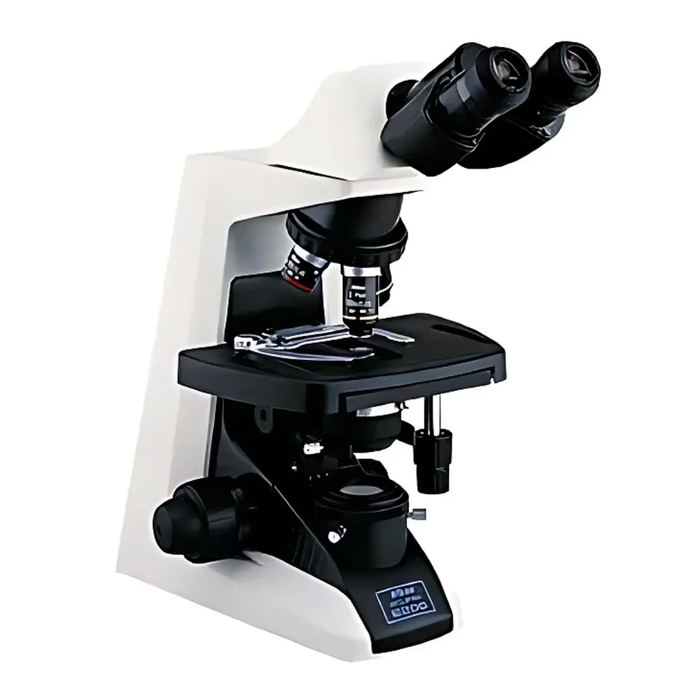
Nikon E200 Biological Microscope
| Brand | Nikon |
|---|---|
| Origin | Japan |
| Microscope Type | Upright Biological Microscope |
| Model | E200 |
| Optical System | CFI60 Infinity-Corrected |
| Magnification Range | 40×–1000× |
| Eyepiece Tube | E2-TB Binocular (hinged, 30° inclination) or E2-TF Trinocular (30° inclination, 360° rotation, interpupillary distance 47–75 mm) |
| Eyepieces | CFI E10×/20 mm (field number 20 mm), optional CFI 15×/12 mm |
| Objectives | CFI E Plan Achromat 4× (NA 0.10), 10× (NA 0.25), 40× (NA 0.65), 100× oil (NA 1.25) |
| Nosepiece | Internal quadruple revolving nosepiece |
| Stage | Rectangular mechanical stage (216 × 150 mm), X–Y travel 78 × 54 mm, low-position coaxial controls, auto-return focusing mechanism |
| Condenser | E2 Abbe condenser (NA 1.25) with iris diaphragm and objective position markers |
| Illumination | 6 V / 20 W halogen lamp, centerable and height-adjustable |
| Focus Mechanism | Coarse focus (37.7 mm per revolution), fine focus (0.2 mm per revolution, 2 µm graduation), adjustable coarse torque, built-in stage stop for high-magnification protection |
| Compliance | Designed for ISO 10993-5 biocompatibility of user-contact components |
Overview
The Nikon E200 Biological Microscope is an upright, infinity-corrected optical instrument engineered for routine laboratory applications in life science education, clinical diagnostics, and basic research. Built upon Nikon’s CFI60 optical architecture—the first generation of Nikon’s 60 mm parfocal distance infinity-corrected system—the E200 delivers consistent image fidelity across its full magnification range (40× to 1000×). Its optical design minimizes chromatic and spherical aberrations through plan achromat objectives and wide-field eyepieces, enabling reliable morphological assessment of stained tissue sections, microbial cultures, blood smears, and fixed cell preparations. The microscope adheres to fundamental ergonomic principles defined by ISO 9241-5 and IEC 62366-1, ensuring operator comfort during extended observation sessions without compromising mechanical stability or optical repeatability.
Key Features
- CFI60 infinity-corrected optical pathway ensures uniform resolution and flatness of field across all magnifications, supporting future upgrades with contrast-enhancing accessories.
- Integrated auto-return mechanical stage: Pressing the stage downward releases tension for rapid slide exchange; release restores original focal plane—eliminating manual re-focusing between specimens.
- Hinged binocular or trinocular tube (E2-TB/E2-TF) with 30° inclination and 360° rotation enables natural neck posture and accommodates varied user anthropometry; interpupillary adjustment range (47–75 mm) supports broad user demographics.
- Low-profile coaxial X–Y stage controls positioned at optimal reach distance reduce shoulder strain; stage surface (216 × 150 mm) accommodates dual slides simultaneously.
- Robust monolithic frame design embeds vertical focusing rails (188.5 mm travel length) within the base housing, enhancing torsional rigidity and vibration damping—critical for stable high-NA oil-immersion imaging.
- Halogen illumination system (6 V / 20 W) features centerable condenser and calibrated iris diaphragm with engraved NA alignment markers corresponding to each objective lens—ensuring Köhler illumination consistency per ISO 8578.
Sample Compatibility & Compliance
The E200 is optimized for transmitted-light brightfield microscopy of standard glass-mounted biological specimens: histological sections (paraffin-embedded or frozen), cytological smears, microbiological preparations (Gram-stained bacteria, fungal elements), and hematology slides. It accepts standard 1 mm-thick microscope slides (76 × 26 mm) and No. 1.5 cover glasses (0.17 mm thickness). Optional accessories—including phase contrast sliders, darkfield condensers, dual- and quad-band fluorescence cubes, and polarizing filters—extend compatibility to unstained live-cell observation, birefringent material analysis, and basic fluorescence screening. The instrument complies with IEC 61010-1 safety standards for laboratory electrical equipment and incorporates anti-fungal treatment on ocular lenses and rubber eyecups per ISO 846 (method C). While not FDA-cleared as a medical device, it meets functional requirements outlined in CLIA-waived testing environments when used with validated staining protocols.
Software & Data Management
The E200 operates as a standalone optical platform but integrates seamlessly with third-party digital imaging systems via its trinocular port (22.5 mm C-mount thread). When paired with compliant CMOS cameras (e.g., Nikon DS-Fi3, or certified USB3.0 models), it supports DICOM-compliant image capture, timestamped metadata logging (including objective ID, magnification, illumination intensity), and audit-trail-enabled export—facilitating alignment with 21 CFR Part 11 requirements where electronic records are employed. Nikon NIS-Elements Basic software (sold separately) provides measurement tools (calibrated linear, area, and angle functions), multi-focus stacking, and annotation layers compatible with institutional LIMS interfaces. All firmware updates are distributed via Nikon’s secure enterprise portal with SHA-256 signature verification.
Applications
- Undergraduate & graduate teaching: Standardized curriculum delivery in histology, microbiology, and cell biology labs—leveraging consistent magnification steps, intuitive controls, and durability under high-throughput student use.
- Clinical pathology support: Routine examination of H&E-stained biopsy sections, Pap smears, and peripheral blood films in hospital satellite labs operating under CAP-accredited quality management systems.
- Quality control in biomanufacturing: Visual inspection of cell culture morphology pre-harvest, sterility checks of media filtration units, and verification of mycoplasma contamination using 100× oil immersion.
- Botanical & zoological taxonomy: Morphometric analysis of pollen grains, diatom frustules, and insect cuticle structures requiring high-contrast brightfield visualization.
- Veterinary diagnostics: Examination of skin scrapings, ear swabs, and fecal flotation preparations in companion animal practice settings.
FAQ
Is the Nikon E200 compatible with digital camera systems?
Yes—the trinocular version (E2-TF) features a standardized 22.5 mm C-mount interface and supports both analog video output and direct USB3.0 camera integration. Third-party SDKs enable custom API development for automated image acquisition pipelines.
Does the E200 meet regulatory requirements for GLP or GMP environments?
While the base instrument lacks embedded electronic record functionality, its mechanical reproducibility, calibration traceability (via optional stage micrometer), and compatibility with 21 CFR Part 11–compliant imaging software make it suitable for regulated workflows when deployed within documented SOPs.
Can phase contrast or fluorescence be added post-purchase?
Yes—Nikon offers factory-aligned phase contrast sliders (PH1–PH3), LED-based fluorescence illuminators (integrated or external), and dichroic filter sets validated for FITC/TRITC/DAPI excitation bands. Installation requires no optical realignment.
What maintenance intervals are recommended for the halogen illumination system?
Lamp replacement is typically required every 80–100 hours of operation. Nikon recommends quarterly cleaning of condenser lenses and annual verification of Köhler alignment using the included centering telescope—procedures detailed in the service manual (PN: E200-SM-EN).
Is the auto-return stage mechanism serviceable in the field?
The spring-loaded return mechanism is sealed and non-user-serviceable; however, Nikon-certified technicians can replace the entire stage assembly (P/N: E2-STG-ASSY) with ≤2-hour downtime under standard warranty coverage.
Related Products


