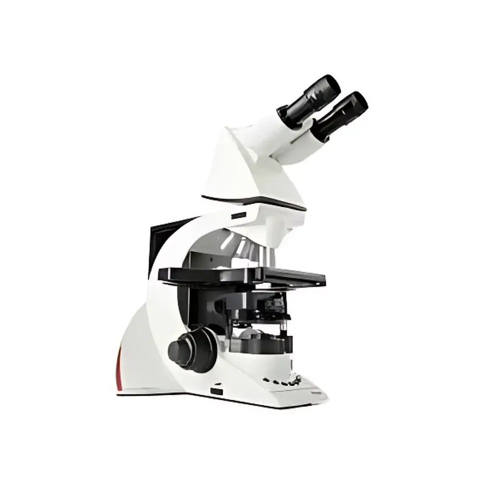
Leica DM3000 Biological Microscope
| Brand | Leica |
|---|---|
| Origin | Italy |
| Model | DM3000 |
| Type | Upright Biological Microscope |
| Automation | Motorized Objective Turret (6-position), Motorized Condenser Head, Auto Illumination Control |
| Ergonomics | Adjustable-Height Focus Knobs (Patent DE10340721), Symmetrical XY Stage & Focus Controls, ErgoStage with Ceramic Surface |
| Optical System | HC PLAN S Widefield Eyepieces (10×/22 mm), HI PLAN Achromatic & Phase Contrast Objectives (4×, 10×, 20×, 40×, 100× Oil), Integrated Daylight Filter (32 mm), Halogen Illumination (12 V / 30 W) |
| Software Compatibility | Leica Application Suite (LAS) for Pathology & Cytology Imaging |
| Compliance | Designed to support GLP/GMP workflows, ISO 13485-aligned manufacturing, CE-marked per EU Medical Device Regulation (MDR 2017/745) for in vitro diagnostic support applications |
Overview
The Leica DM3000 is a high-performance upright biological microscope engineered for routine and advanced applications in clinical pathology, cytology, hematology, and biomedical research laboratories. Built upon Leica Microsystems’ decades of optical precision engineering, the DM3000 employs a modular, ergonomically optimized platform centered on reproducible Köhler illumination, parfocal and parcentric optics, and intelligent motorized automation. Its core optical architecture complies with DIN/ISO 8036 standards for objective labeling, magnification fidelity, and mechanical tube length consistency (160 mm finite system). The instrument integrates a stable, vibration-damped stand with a rigid optical path designed to minimize thermal drift and maintain focus stability over extended imaging sessions — a critical requirement for slide-based diagnostic workflows where inter-operator consistency directly impacts diagnostic confidence.
Key Features
- Motorized 6-position objective turret with sub-second switching (<500 ms) between user-defined objectives; programmable via front-panel buttons or optional footswitch
- Motorized condenser head (UCA/P type) that automatically positions the aperture diaphragm and field diaphragm at optimal settings for each magnification — ensuring consistent contrast and resolution across 4× to 100× oil immersion
- Auto-illumination control: real-time adjustment of halogen lamp intensity based on selected objective’s transmission characteristics and numerical aperture — eliminating manual brightness recalibration during multi-magnification review
- Ergonomic design certified under ISO 9241-5: adjustable-height focus knobs (patented DE10340721), symmetrical XY-stage and focus controls, 15° inclined trinocular tube, and ceramic-surfaced ErgoStage for wear resistance and smooth specimen navigation
- Modular compatibility: supports Leica HC PLAN S widefield eyepieces (10×/22 mm field number), phase contrast accessories (PH1–PH3), fluorescence filter cubes (optional), and LAS-compatible digital camera interfaces
- Programmable control interface: all physical buttons — including optional footswitch inputs — are fully customizable for functions such as illumination toggle, objective recall, fluorescence mode activation, or LAS software triggering
Sample Compatibility & Compliance
The Leica DM3000 accommodates standard 1″ × 3″ (25 × 75 mm) glass microscope slides and accommodates thicknesses from 0.13–1.2 mm, including coverslip-corrected preparations (0.17 mm coverslip specification). Its condenser system (UCA/P, NA 1.6) supports brightfield, phase contrast (with dedicated annuli sets), and optional fluorescence modalities using Leica’s high-transmission filter sets. The system meets IEC 61000-6-3 (EMC emissions) and IEC 61000-6-2 (immunity) standards. As a Class I medical device per EU MDR 2017/745, it is intended for in vitro diagnostic support — particularly in histopathology and cytopathology workflows compliant with CAP, CLIA, and ISO 15189 requirements. Its audit-ready operation supports GLP/GMP documentation when paired with Leica LAS X software featuring 21 CFR Part 11-compliant electronic signatures and full audit trail functionality.
Software & Data Management
The DM3000 is natively compatible with Leica Application Suite (LAS) and LAS X platforms, enabling seamless integration into digital pathology pipelines. LAS X Core provides synchronized image acquisition, annotation, measurement (area, length, particle count), and report generation aligned with DICOM-SR and HL7 messaging standards. When configured with a Leica DFC camera, the system supports live Z-stack acquisition, multi-channel fluorescence overlay, and time-lapse capture — all with hardware-triggered synchronization to eliminate motion artifacts. All metadata (objective ID, magnification, illumination intensity, condenser position, date/time stamp) is embedded in TIFF and JPEG2000 exports. For enterprise deployment, LAS X Enterprise supports centralized license management, role-based access control, and PACS integration via DICOM Modality Worklist (MWL) and Storage Commitment protocols.
Applications
The DM3000 serves as a primary diagnostic tool in clinical cytology labs performing Pap smears, urine sediment analysis, and fine-needle aspiration evaluations. In surgical pathology, it supports rapid intraoperative frozen section assessment and permanent slide triage. Its phase contrast capability enables label-free live-cell observation in academic and pharmaceutical research — particularly for hematopoietic cell morphology, stem cell differentiation assays, and microbiological identification (e.g., acid-fast bacilli staining). The motorized workflow reduces repetitive strain injury (RSI) risk during high-volume screening, while its standardized optical calibration ensures inter-laboratory comparability — a prerequisite for multicenter clinical trials requiring morphometric consistency.
FAQ
Is the Leica DM3000 compliant with FDA 21 CFR Part 11 for electronic records?
Yes — when operated with Leica LAS X software configured in “Audit Trail Mode”, the system enforces user authentication, electronic signatures, and immutable activity logging required under Part 11 for regulated environments.
Can the DM3000 be upgraded to support fluorescence imaging?
Yes — the base stand includes a fluorescence-ready light path; users may add Leica filter cubes (e.g., GFP, TRITC, DAPI), a mercury or LED excitation source, and appropriate emission filters without hardware modification.
What maintenance is required for the motorized components?
Leica recommends annual calibration of turret positioning accuracy and condenser alignment using the included focusing telescope; no routine lubrication or internal servicing is required under normal laboratory use conditions.
Does the DM3000 support third-party camera systems?
Yes — via C-mount adapter (1× or 0.5× reduction), the trinocular port supports industry-standard USB3/UVC and GigE cameras from manufacturers including Basler, Hamamatsu, and FLIR, provided driver compatibility is verified through Leica’s hardware interoperability matrix.
How does the auto-illumination system ensure photostability during long acquisitions?
The system dynamically modulates lamp voltage rather than using neutral density filters, minimizing thermal load on specimens while maintaining constant photon flux density at the sample plane — critical for reducing photobleaching in fluorescent preparations.


