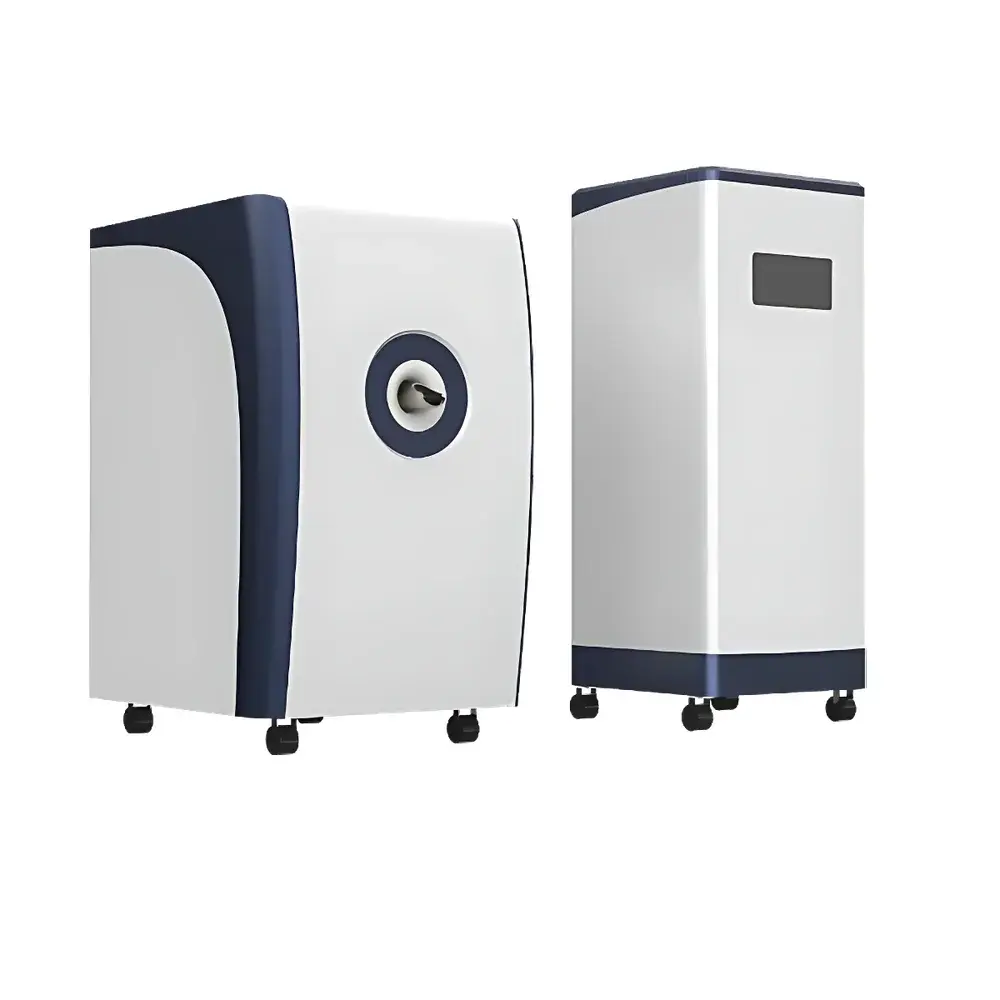
HTNMR HT-MRSI60-35 1.5T Small Animal MRI System with 35 mm Bore
| Brand | HTNMR |
|---|---|
| Origin | Shanghai, China |
| Manufacturer Type | OEM Manufacturer |
| Region of Origin | Domestic (China) |
| Model | HT-MRSI60-35 |
| Instrument Type | Magnetic Resonance Imaging (MRI) System |
| Animal Species | Mouse (also compatible with rat and small plant specimens) |
| Maximum Sample Diameter | 35 mm |
| Maximum Sample Height | 50 mm |
| Spatial Resolution | 0.08 mm (high-resolution mode) |
| Field Homogeneity | ≤ 8 ppm over DSV 20 mm |
| Field Stability | Frequency drift ≤ 100 Hz/h |
| Imaging Matrix Options | 128×128×128 (standard), 256×256×128 (high-res), 512×512×128 (ultra-high-res) |
| Multi-nuclear Capability | ¹H, ¹⁹F, ³¹P |
| Magnet Type | Permanent Magnet (1.5 T) |
| Cryogen-Free | Yes |
| Passive Shielding | Integrated self-shielding design |
| RF Coil Configuration | Tunable multi-nuclei volume coil (standard) |
| Number of Simultaneous Samples | 1 |
Overview
The HTNMR HT-MRSI60-35 is a compact, cryogen-free 1.5 Tesla permanent-magnet small animal MRI system engineered for preclinical research in neuroscience, oncology, cardiology, and molecular imaging. Unlike superconducting systems requiring liquid helium and active shielding, this platform leverages a high-stability permanent magnet architecture—delivering consistent 1.5 T field strength within a 35 mm clear bore while eliminating cryogenic infrastructure, magnetic fringe field mitigation requirements, and associated operational overhead. The system operates on the fundamental principle of nuclear magnetic resonance: hydrogen (¹H), fluorine (¹⁹F), and phosphorus (³¹P) nuclei align under a strong static magnetic field; radiofrequency (RF) pulses perturb this alignment, and the resulting relaxation signals (T₁, T₂, T₂*) are spatially encoded using gradient fields and reconstructed into quantitative 2D/3D anatomical or functional images. Designed specifically for murine models—including mice and rats—the HT-MRSI60-35 supports longitudinal in vivo studies under controlled physiological conditions, with integrated physiological monitoring interfaces (optional) compatible with respiratory gating and ECG synchronization.
Key Features
- Permanent-magnet 1.5 T field generation with intrinsic thermal stability and <100 Hz/h frequency drift—enabling reproducible longitudinal acquisitions without real-time shimming recalibration.
- Integrated self-shielding design eliminates need for external RF cages or passive ferromagnetic shielding rooms—reducing facility footprint and installation complexity.
- Multi-nuclear RF capability (¹H/¹⁹F/³¹P) supported by broadband tunable volume coils, facilitating dual-contrast agent validation, metabolic imaging (e.g., ³¹P spectroscopy), and fluorinated tracer tracking.
- High-fidelity 3D imaging up to 512 × 512 × 128 matrix resolution with isotropic voxel dimensions down to 0.08 mm—achievable across standard spin-echo, gradient-echo, inversion-recovery, and fast spin-echo sequences.
- Uniformity optimized to ≤8 ppm over a 20 mm diameter spherical volume (DSV), ensuring quantitative accuracy in relaxation time mapping and contrast agent pharmacokinetic modeling.
- Open-access pulse sequence framework allows user-defined sequence development via MATLAB-based SDK—supporting advanced diffusion-weighted imaging (DWI), arterial spin labeling (ASL), and dynamic contrast-enhanced (DCE) protocols.
Sample Compatibility & Compliance
The HT-MRSI60-35 accommodates live mice (up to ~35 g), juvenile rats (up to ~250 g), excised organs, and small botanical specimens within its 35 mm × 50 mm cylindrical field-of-view. Anesthesia integration (isoflurane-compatible gas delivery ports) and temperature-regulated animal bed (37 °C ± 0.5 °C) ensure physiological stability during scans lasting up to 90 minutes. All hardware and firmware comply with IEC 61000-6-3 (EMC emission standards) and IEC 61000-6-4 (industrial immunity). Data acquisition workflows support audit-trail logging and electronic signatures per FDA 21 CFR Part 11 when paired with validated LIMS or ELN integrations—making the system suitable for GLP-compliant toxicology and regulatory submission studies.
Software & Data Management
The system runs on HTNMR’s proprietary MR Studio v4.2 software suite, built on a DICOM 3.0–compliant architecture with native support for NIfTI and Analyze file formats. Image reconstruction, post-processing (including ROI-based T₁/T₂ fitting, ADC map generation, and co-registration with optical or CT data), and batch analysis are performed within a scriptable Python API environment. Raw k-space data export enables third-party reconstruction (e.g., Berkeley Advanced Reconstruction Toolbox, BART) and machine learning pipeline integration. All datasets include embedded metadata tags compliant with MINC and BIDS specifications—ensuring interoperability with public repositories such as OpenNeuro and Preclinical Imaging Data Repository (PIDR).
Applications
- Longitudinal tumor growth monitoring and anti-angiogenic therapy assessment via dynamic contrast-enhanced MRI (DCE-MRI) and vessel size index (VSI) mapping.
- Cardiac functional analysis in transgenic mouse models—including cine-MRI for ejection fraction, wall motion, and diastolic strain rate quantification.
- Molecular imaging of inflammation using ¹⁹F-labeled perfluorocarbon nanoemulsions and targeted ³¹P-phospholipid probes.
- Neuroanatomical phenotyping in neurodegenerative models (e.g., APP/PS1 mice) via high-resolution 3D T₂-weighted whole-brain morphometry.
- In vitro relaxivity characterization (r₁/r₂) of novel contrast agents under standardized phantom conditions—traceable to NIST SRM 1916a (Gd-DTPA reference).
- Plant root architecture and water transport dynamics imaging in Arabidopsis thaliana and Zea mays seedlings.
FAQ
Is the system compatible with existing preclinical imaging workflows?
Yes—the HT-MRSI60-35 supports DICOM export and integrates with PACS, PMOD, and VivoQuant via standard network protocols (DICOM SCU/SCP, FTP, REST API).
What level of technical support and service coverage is provided?
HTNMR offers on-site installation, operator training, and remote diagnostics through certified field engineers. Extended warranty and preventive maintenance contracts include annual field homogeneity verification and RF coil calibration.
Can the system be upgraded to support advanced sequences like diffusion tensor imaging (DTI)?
Yes—DTI, ASL, and MR spectroscopy (MRS) packages are available as optional software modules, validated against ACR MRI phantom test procedures.
Does the permanent magnet require periodic re-magnetization?
No—HTNMR’s sintered NdFeB magnet assembly is rated for >15 years of continuous operation at 1.5 T with <0.01% field decay per annum under ambient temperature control (20–25 °C).
How is data integrity ensured during long-duration acquisitions?
All acquisitions log timestamped system logs, gradient waveform traces, and RF power monitors. Raw k-space data includes checksums and header-level CRC validation—enabling full traceability from acquisition to publication-grade figure generation.