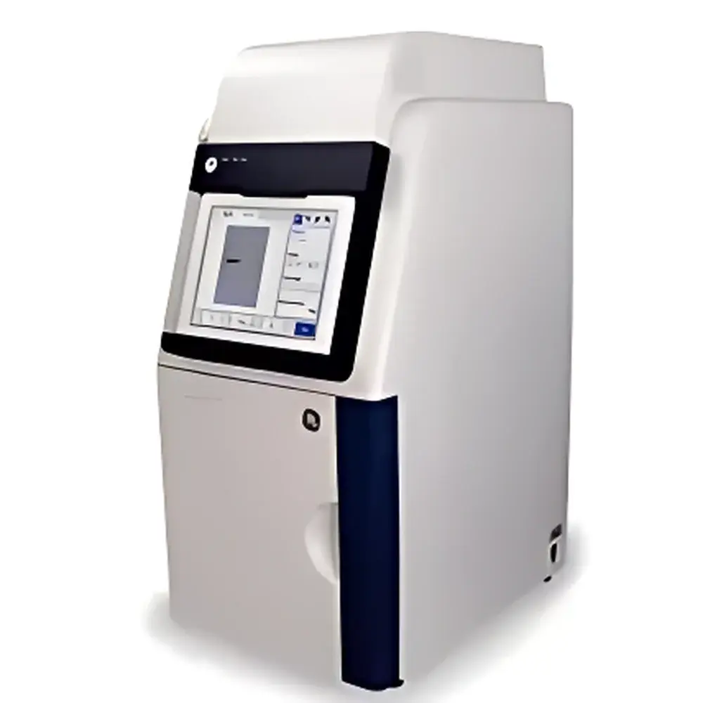
Amersham Imager 600 Series Ultra-Sensitive Multimodal Gel Imaging System
| Brand | Cytiva |
|---|---|
| Origin | USA |
| Model | Amersham Imager 600 |
| Instrument Type | Chemiluminescence Gel Imaging System |
| CCD Resolution | 5.8 MP |
| Bit Depth | 16-bit |
| Dynamic Range | 0–4.8 OD |
| CCD Sensor Size | 15.6 × 23.4 mm |
| Detection Sensitivity | ≥0.02 ng protein, ≥0.02 ng DNA |
| Signal-to-Noise Ratio | ≥56 dB |
| Lens Focal Length | 8–48 mm |
Overview
The Amersham Imager 600 Series is a high-performance, ultra-sensitive multimodal gel imaging system engineered for quantitative detection and analysis of biomolecules across chemiluminescent, fluorescent, UV-transilluminated, and white-light modalities. Designed for rigorous life science laboratories, it employs a scientific-grade cooled CCD sensor with thermoelectric stabilization to minimize dark current noise—enabling reproducible, low-background acquisition essential for low-abundance target detection. Unlike conventional systems relying on fixed-gain or manual exposure tuning, the Imager 600 integrates hardware-level optical optimization—including an f/0.74 fixed-focus lens assembly with high-transmission optical glass—to maximize photon capture efficiency across visible and near-UV wavelengths (254 nm–700 nm). Its architecture supports standardized workflows aligned with GLP-compliant documentation requirements, including timestamped metadata embedding and audit-ready image provenance tracking.
Key Features
- Scientific-grade 5.8-megapixel CCD sensor with 16-bit digitization, delivering linear dynamic range up to 4.8 optical density (OD) units—enabling quantitation across >4 orders of magnitude intensity without saturation.
- Cooled CCD operation at −25°C (typical), reducing thermal noise and supporting long-exposure chemiluminescence imaging with signal stability <±1.5% over 30 min.
- Motorized 8–48 mm zoom lens with calibrated focus positioning and auto-aperture control—ensuring consistent magnification and illumination uniformity across sample formats (gels, blots, microplates, Petri dishes).
- Dual-mode excitation capability: integrated UV transilluminator (254 nm, 302 nm optional) and six-channel LED fluorescence module (365 nm, 470 nm, 530 nm, 590 nm, 630 nm, 670 nm) with user-selectable bandpass filters via side-access drawer.
- 12.1-inch capacitive touchscreen interface with glove-compatible operation, preloaded with ImageQuant TL software for real-time preview, region-of-interest (ROI) definition, background subtraction, and molecular weight calibration.
Sample Compatibility & Compliance
The Amersham Imager 600 accommodates standard electrophoretic formats including mini- and midi-gels (up to 16 × 20 cm), nitrocellulose/PVDF membranes (up to 20 × 25 cm), 96-well and 384-well microplates, and 60–100 mm Petri dishes. All optical paths comply with IEC 61000-4-3 (EMC immunity) and UL 61010-1 safety standards. The system supports ISO/IEC 17025-aligned validation protocols and includes built-in tools for IQ/OQ documentation. ImageQuant TL software meets FDA 21 CFR Part 11 requirements for electronic records and signatures when deployed with network authentication and audit trail configuration.
Software & Data Management
ImageQuant TL v8.4 provides full local and remote control via Ethernet or Wi-Fi, enabling instrument status monitoring, scheduled acquisitions, and live-streaming of preview frames to desktop or mobile clients. Raw images are saved in TIFF format with embedded EXIF metadata (exposure time, gain, lens position, temperature, date/time stamp). Batch processing supports normalization against internal loading controls, densitometric quantitation using cubic spline interpolation, and export to CSV, PDF, or publication-ready PNG/JPEG. Data integrity safeguards include automatic checksum generation, folder-based versioning, and integration with LIMS via RESTful API endpoints.
Applications
- Western blot quantitation of phosphorylated proteins and low-expression targets under chemiluminescent detection (ECL Prime, SuperSignal West Femto).
- High-resolution DNA/RNA gel documentation with ethidium bromide, SYBR Safe, or GelRed staining—including fragment sizing and relative band intensity analysis.
- Fluorescent multiplex imaging of Cy3/Cy5-labeled arrays, GFP/RFP-tagged cell lysates, and quantum dot-conjugated antibodies.
- UV-induced DNA damage assessment via thymine dimer detection using 254 nm transillumination.
- Colony counting and confluence analysis in cell culture assays using white-light reflectance mode.
FAQ
What is the minimum detectable mass for a Coomassie-stained protein band?
Under optimized white-light conditions with 120-second exposure and background subtraction, the system reliably detects ≥2 ng of BSA per band on 1 mm-thick SDS-PAGE gels.
Does the system support time-lapse chemiluminescence imaging?
Yes—programmable multi-frame acquisition allows sequential exposures from 0.1 s to 60 min, with automated alignment and drift correction enabled via fiducial marker tracking.
Can third-party filter sets be installed?
Yes—the side-access filter drawer accepts standard 25 mm diameter threaded filters (M25.5×0.5); custom dichroic and emission filters may be validated for compatibility upon request.
Is remote software update supported?
Firmware and software updates are delivered via secure HTTPS push through Cytiva’s authorized service portal; updates require administrator credentials and generate post-installation verification logs.
How is calibration maintained across instruments?
Each unit ships with NIST-traceable neutral density step tablets and fluorescent reference standards; routine calibration checks are guided by ImageQuant TL’s integrated QC wizard and documented in instrument logbooks.
Related Products


