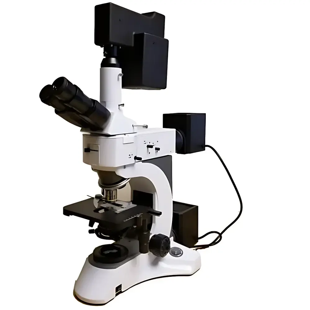
Auniontech Model 4200M Microscopic Staring Hyperspectral Imaging System
| Brand | Auniontech |
|---|---|
| Origin | Imported |
| Manufacturer Type | Authorized Distributor |
| Imaging Principle | Staring (Snapshot) Interferometric Hyperspectral Imaging |
| Spectral Range | 400–1000 nm |
| Spectral Resolution | 4 nm |
| Spectral Channels | 300–600 |
| Spatial Resolution | 1280 × 1024 pixels (2.3 MP) |
| Field of View (TFOV) | Configurable via microscope objective |
| Instantaneous Field of View (IFOV) | Objective-dependent |
| Frame Rate | Up to 30 fps (full cube, dependent on spectral binning) |
| Bit Depth | 8 or 16 bit |
| Interface | USB 3.0 |
| Operating Temperature | 20 °C ± 5 °C |
| Relative Humidity | ≤65% non-condensing |
| Power Supply | 18 VDC (optical head only) |
| Dimensions & Weight | 230 × 120 × 200 mm, 1.4 kg |
Overview
The Auniontech Model 4200M Microscopic Staring Hyperspectral Imaging System is a modular, interferometer-based hyperspectral add-on module engineered for integration with standard upright and inverted optical microscopes. Unlike scanning-based (push-broom or whiskbroom) or filter-wheel-driven multispectral systems, the Model 4200M employs a spatially resolved, snapshot Fourier-transform interferometric architecture—enabling simultaneous acquisition of full spectral data cubes (λ × x × y) at every pixel without mechanical scanning or temporal multiplexing. This staring modality ensures high photon efficiency, intrinsic spatial-spectral registration stability, and immunity to motion artifacts—critical for live-cell imaging, time-lapse tissue analysis, and quantitative microspectroscopy. The system operates across the visible to near-infrared (VIS-NIR) range (400–1000 nm), delivering up to 600 spectrally resolved channels with a nominal resolution of 4 nm FWHM. Its compact form factor (230 × 120 × 200 mm, 1.4 kg) and USB 3.0 interface allow seamless deployment in regulated laboratory environments, including ISO 17025-accredited analytical labs and GLP-compliant preclinical research facilities.
Key Features
- Staring interferometric design: Eliminates point-scanning or line-scanning mechanisms—preserves native microscope resolution (no CFA-induced spatial aliasing or pixel binning)
- Programmable spectral sub-band acquisition: Users define custom wavelength ranges and channel spacing via software API; avoids redundant data capture and reduces storage/compute overhead
- Native 2.3 MP spatial sampling (1280 × 1024) with objective-coupled IFOV scalability: Supports 10× to 100× microscope objectives without optical relaying loss
- Dual-bit-depth operation (8/16 bit): Enables dynamic range optimization for low-light fluorescence or high-intensity reflectance applications
- Thermally stabilized optical head: Maintains interferometric path-length consistency within ±0.5 µm over 20 °C ± 5 °C operating range
- USB 3.0 real-time streaming: Sustains full-cube frame rates up to 30 fps with on-the-fly spectral binning (e.g., 150-channel mode at 60 fps)
Sample Compatibility & Compliance
The Model 4200M is compatible with standard transmitted-light and epi-illumination microscope configurations—including brightfield, phase contrast, DIC, and fluorescence modalities. It supports both fixed and live biological specimens (e.g., histological sections, cell monolayers, organoids), thin-film materials (polymers, semiconductors, coatings), and forensic trace evidence (ink, fibers, gunshot residue). Data output conforms to HDF5 and ENVI-compatible formats, ensuring interoperability with third-party spectral unmixing tools (e.g., ENVI, MATLAB Hypercube Toolbox). The system meets CE marking requirements for electromagnetic compatibility (EN 61326-1) and safety (EN 61010-1). While not FDA-cleared as a medical device, its spectral fidelity and repeatability support method validation per ASTM E1777 (Standard Practice for Hyperspectral Imaging Data Acquisition) and ISO/IEC 17025 clause 7.2.2 (validation of non-standard methods).
Software & Data Management
The included Auniontech Hyperspectral Studio v4.x provides a complete workflow: hardware control, real-time preview, spectral calibration (using NIST-traceable tungsten-halogen and mercury-argon sources), radiometric correction, and endmember extraction via vertex component analysis (VCA) or N-FINDR. All acquisitions include embedded metadata (wavelength grid, exposure time, objective magnification, illumination type) compliant with the MIAPPE 1.1 specification for reproducible imaging experiments. Audit trails record user actions, parameter changes, and processing steps—supporting 21 CFR Part 11 compliance when deployed with optional electronic signature modules. Raw data cubes are stored in lossless compression (HDF5 + LZ4), with optional export to CSV, TIFF stacks, or MxN spectral matrices for integration into Python (scikit-learn, hylite) or R (hyperSpec) analytics pipelines.
Applications
- Life Sciences: Multiplexed fluorescence unmixing of >5 spectrally overlapping dyes (e.g., FITC/TRITC/Cy5) using single-excitation broadband illumination; quantification of NAD(P)H/FAD redox ratios in metabolic phenotyping
- Pharmaceutical QA: Spatial mapping of active pharmaceutical ingredient (API) distribution in tablet cross-sections; detection of coating thickness variation and excipient segregation
- Forensics: Non-destructive differentiation of ink formulations on questioned documents; identification of accelerant residues via C–H and O–H vibrational overtone signatures in NIR
- Materials Science: Mapping of crystallinity gradients in polymer blends; defect localization in perovskite thin films via photoluminescence peak shift analysis
- Agriculture & Food: Early detection of fungal infection in grain kernels via chlorophyll fluorescence quenching; classification of olive oil adulteration using lipid absorption features at 970 nm and 1210 nm
- Cultural Heritage: Pigment identification in historical manuscripts without sampling; monitoring of varnish degradation kinetics through carbonyl band evolution at 1720 cm⁻¹ (converted from NIR reflectance)
FAQ
Does the Model 4200M require external calibration sources?
Yes—routine calibration uses supplied NIST-traceable tungsten-halogen (radiometric) and mercury-argon (wavelength) standards. Factory calibration is performed annually; field recalibration takes <5 minutes.
Can it be integrated with automated microscope stages?
Yes—via TTL trigger input/output and RS-232/USB serial commands. Compatible with Prior, Ludl, and Marzhauser motorized stage controllers.
Is spectral data export compatible with MATLAB or Python?
Yes—raw .h5 files contain fully documented datasets (wavelength vector, intensity cube, metadata) readable via h5py, SciPy, or the open-source HySpex Python library.
What microscope brands and models are certified for coupling?
Validated interfaces exist for Olympus BX series, Nikon Eclipse Ci, Zeiss Axio Imager, and Leica DMi8; custom C-mount or infinity-corrected tube lens adapters available upon request.
Does the system support real-time spectral classification during acquisition?
Yes—user-defined spectral libraries (e.g., mineral spectra from USGS, pigment libraries from FORS database) can be loaded for on-the-fly matched-filter or SAM scoring with latency <200 ms per frame.