BeonChip BE-TRANSFLOW Microfluidic Cell Culture Chip for Complex Porous Membrane-Based Models
| Brand | BEOnChip |
|---|---|
| Origin | Spain |
| Model | BE-TRANSFLOW |
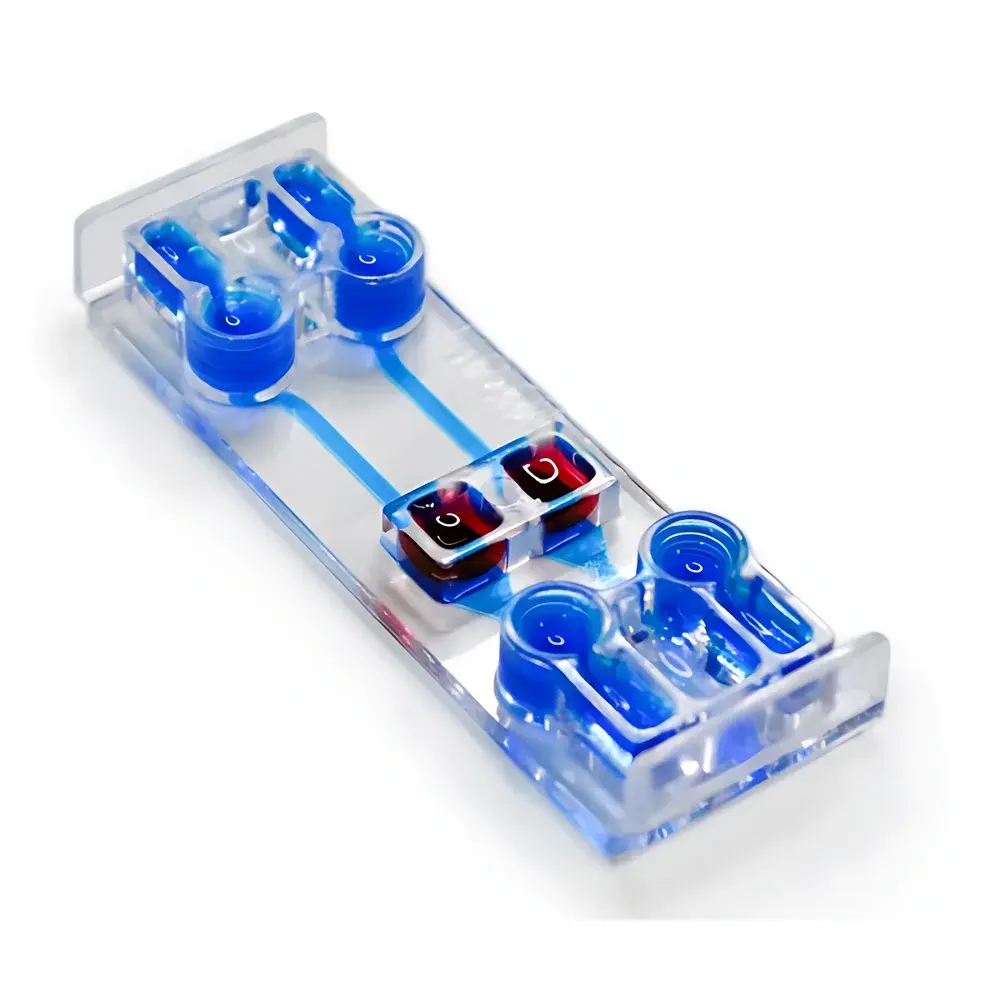
| Form Factor | Standard Microscope Slide (75 × 25 mm) |
| Membrane | Integrated Porous Polycarbonate or PET Membrane (Typical Pore Sizes: 0.4 µm, 3.0 µm, 8.0 µm — Configurable) |
| Chamber Configuration | Dual Independent Culture Wells Connected via Sub-membrane Microchannel Network |
| Optical Compatibility | Fully Transparent, High-Clarity Polymer (Cyclic Olefin Copolymer, COC) |
| Fluidic Interface | Standard Luer Lock Inlets/Outlets (Compatible with Fluigent, Elveflow, Harvard Apparatus, and Other Pressure/Peristaltic/Syringe Pump Systems) |
| Sterility | Pre-sterilized (EO or Gamma), Ready-to-Use |
| Regulatory Status | For Research Use Only (RUO) |
Overview
The BeonChip BE-TRANSFLOW is a standardized, microscope-slide-format microfluidic cell culture chip engineered for advanced in vitro physiological modeling requiring spatially segregated yet fluidically coupled cellular compartments. It operates on the principle of controlled transmembrane mass transport across an integrated porous membrane—enabling precise establishment and maintenance of air–liquid interface (ALI), epithelial/endothelial barrier, and paracrine crosstalk conditions. Unlike conventional Transwell® inserts, the BE-TRANSFLOW integrates microfabricated channel architecture beneath the membrane to support dynamic perfusion, shear stress application, and real-time solute gradient formation. Its monolithic COC construction ensures optical clarity (transmission >92% from 350–700 nm), dimensional stability under prolonged culture (up to 21 days), and minimal non-specific protein adsorption—critical for reproducible immunostaining, live-cell imaging, and quantitative endpoint assays.
Key Features
- Standard 75 × 25 mm footprint—direct compatibility with inverted and upright microscopes, automated slide scanners, and standard incubator trays.
- Dual independent culture wells positioned above a shared, membrane-spanning microchannel network—enabling asymmetric stimulation, directional transport studies, and compartmentalized co-culture.
- Pre-integrated, track-etched polycarbonate or PET membranes (pore sizes configurable per application: 0.4 µm for tight barrier integrity, 3.0 µm for leukocyte transmigration, 8.0 µm for stromal cell infiltration).
- Luer-lock fluidic ports (2 inlet + 2 outlet) designed for low-dead-volume connection to external pump systems—including pressure-driven (Fluigent MFCS-EZ), syringe-based (Harvard PHD Ultra), and peristaltic platforms—ensuring laminar, pulse-free flow at physiological shear rates (0.1–20 dyn/cm²).
- Surface chemistry optimized for direct cell seeding without extracellular matrix pre-coating (optional fibronectin/collagen I coating available for primary endothelial or neuronal cultures).
- Reusable mounting frame option (sold separately) for long-term perfusion experiments requiring repeated media exchange without chip removal.
Sample Compatibility & Compliance
The BE-TRANSFLOW supports primary human cells (e.g., bronchial epithelial cells, brain microvascular endothelial cells, intestinal organoid-derived monolayers), immortalized lines (Caco-2, Calu-3, hCMEC/D3), and 3D co-cultures (epidermal keratinocytes + dermal fibroblasts; osteoblasts + osteoclasts). It has been validated in protocols aligned with ISO 10993-5 (cytotoxicity), OECD TG 431 (skin irritation), and ASTM E2924-22 (microphysiological system characterization). All chips are manufactured under ISO 13485–aligned cleanroom conditions and supplied sterile (ethylene oxide or gamma irradiation), with full traceability documentation provided. Not intended for clinical diagnostic use; labeled “For Research Use Only” per FDA 21 CFR §809.10 and EU IVDR Annex XVI.
Software & Data Management
While the BE-TRANSFLOW is a passive hardware platform, it is fully interoperable with third-party data acquisition ecosystems. When paired with compatible pumps (e.g., Fluigent’s FlowEZ software) or environmental controllers (Okolab stage-top incubators), users can log flow rate, pressure, temperature, and CO₂ in synchronized time-series format. Image data acquired via confocal, phase-contrast, or fluorescence microscopy may be annotated and quantified using open-standard tools (Fiji/ImageJ, Imaris, HALO) with spatial registration to chip landmarks. Audit trails for experimental metadata (cell passage number, membrane lot ID, perfusion duration) are recommended to comply with GLP-aligned recordkeeping practices.
Applications
- Air–liquid interface (ALI) culture of respiratory or corneal epithelia—enabling mucociliary differentiation, aerosol exposure testing, and host–pathogen interaction studies under physiologically relevant oxygen tension.
- Barrier function assessment: Quantitative TEER measurement (via integrated electrodes in optional electrode-equipped variants), FITC-dextran permeability, and immunofluorescent tight junction protein localization (ZO-1, occludin).
- Endothelial–epithelial crosstalk modeling: e.g., blood–brain barrier (BBB) with iPSC-derived brain microvascular endothelial cells and astrocytes; gut–vasculature interface with Caco-2 monolayers and HUVECs.
- Indirect toxicity screening: Paracrine signaling analysis between hepatocyte spheroids and cardiomyocytes across the membrane—avoiding direct contact while preserving soluble mediator exchange.
- Bone-on-a-chip applications: Osteoblast–osteoclast co-culture under mechanical strain (via integrated pneumatic actuators in custom configurations) to model RANKL/OPG-mediated remodeling dynamics.
FAQ
Is the BE-TRANSFLOW compatible with high-resolution live-cell imaging?
Yes—the COC substrate exhibits negligible auto-fluorescence below 500 nm and thermal expansion coefficients matched to glass coverslips, minimizing focus drift during time-lapse acquisition.
Can I reuse the chip after sterilization?
No—BE-TRANSFLOW chips are single-use, pre-sterilized devices. Re-sterilization compromises membrane integrity and surface chemistry.
What pore size should I select for blood–brain barrier modeling?
A 0.4 µm pore size is recommended for restrictive barrier formation with human iPSC-derived endothelial cells; 3.0 µm pores are used when studying immune cell transmigration across the same barrier.
Does BeonChip offer custom membrane coatings or integrated sensors?
Yes—custom ECM coatings (laminin-521, collagen IV), embedded gold microelectrodes for real-time TEER, and PDMS-integrated pressure sensors are available under NRE agreements.
How is the chip mounted for perfusion without leakage?
It uses a standardized gasketed holder (included in Starter Kits) that seals all fluidic interfaces via uniform compression force—validated at pressures up to 150 kPa with no observable leakage over 72-hour runs.


