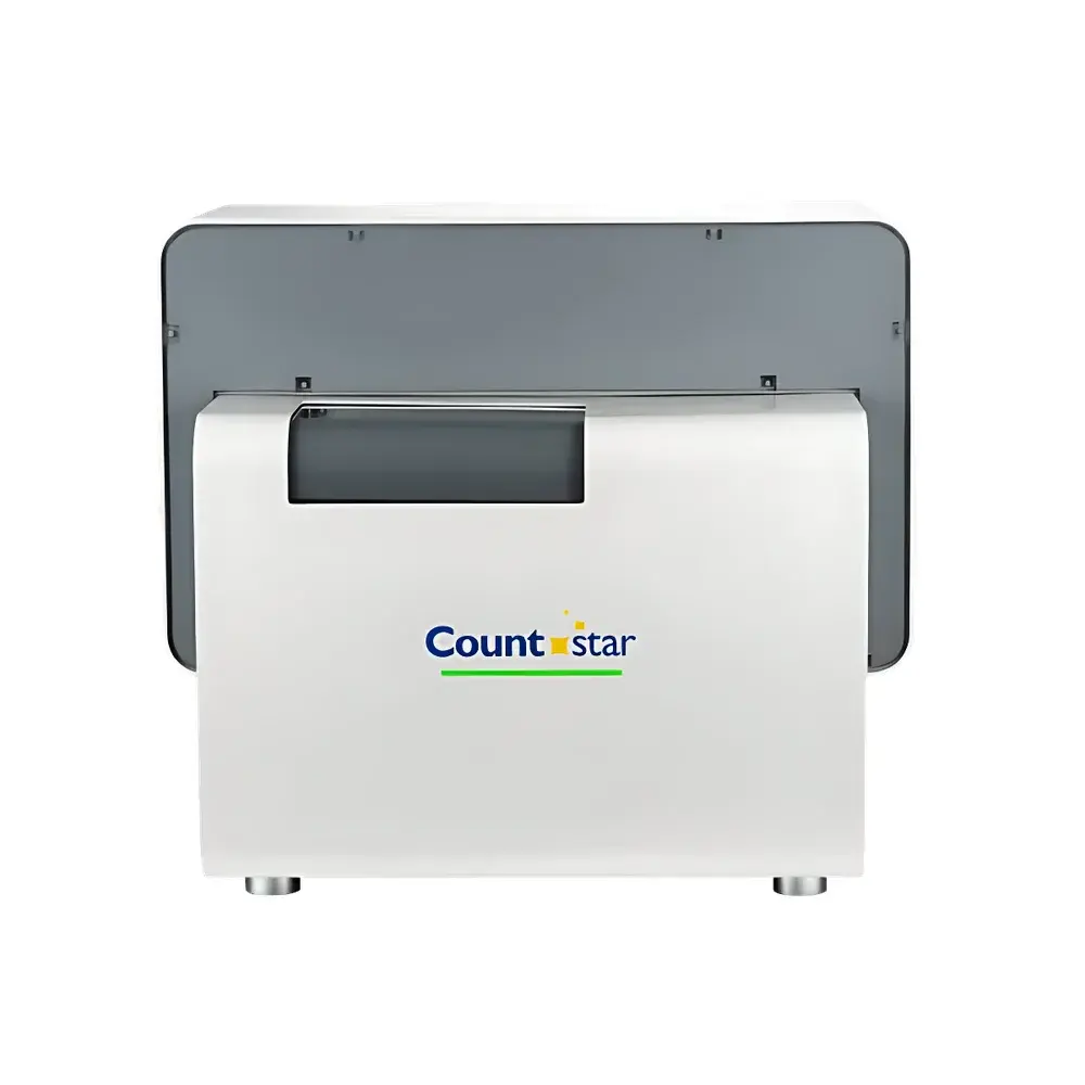
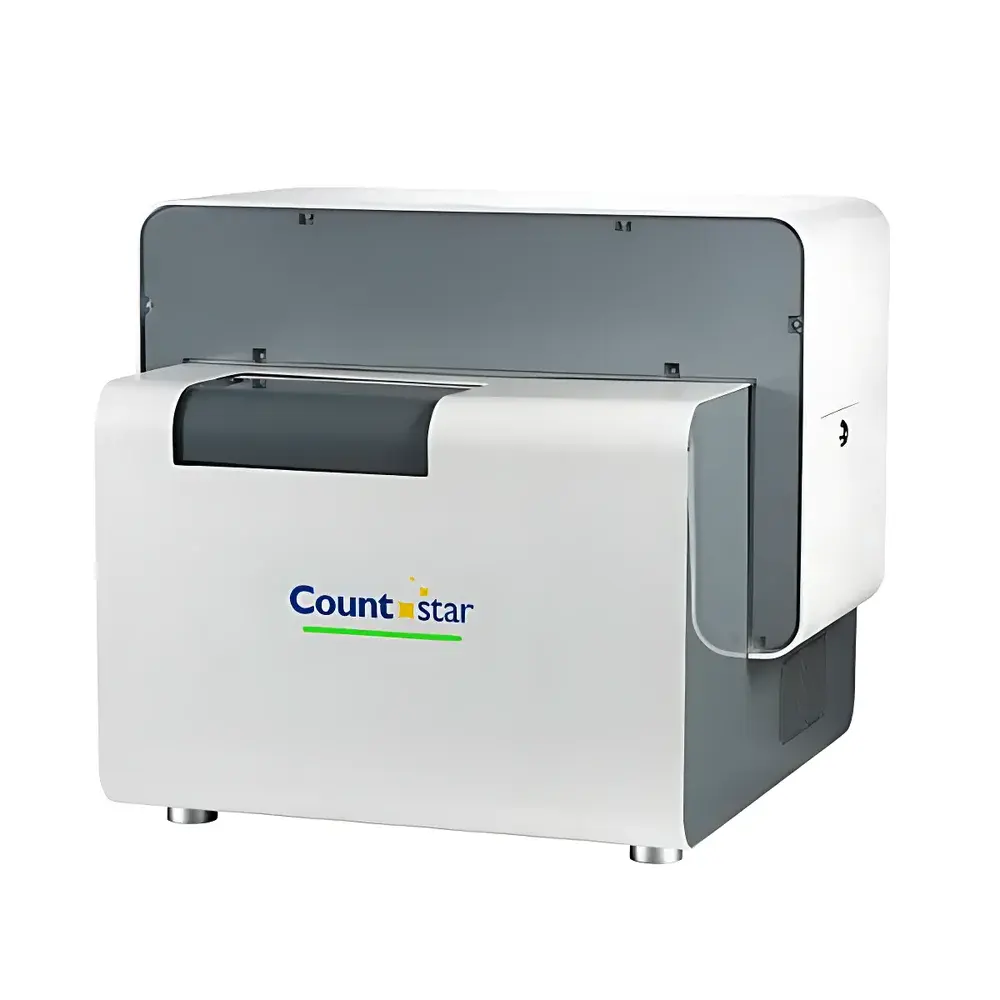
Countstar Castor X High-Throughput Intelligent Cell Imaging and Analysis System
| Brand | Countstar |
|---|---|
| Origin | Shanghai, China |
| Model | Castor X |
| Fluorescence Channels | Dual (Green & Red) |
| Compatible Vessels | 6/12/24/96/384-well plates, Petri dishes, culture flasks |
| Imaging Sensor | Cooled high-resolution monochrome CMOS camera |
| Analysis Engine | AI-powered deep learning algorithms for clonal identification, confluency quantification, and transfection efficiency scoring |
| Software | Modular, GLP-compliant with audit trail and user permission management |
| Regulatory Alignment | Designed to support ISO 13485 workflows and FDA 21 CFR Part 11–ready data integrity practices |
Overview
The Countstar Castor X is a high-throughput intelligent cell imaging and analysis system engineered for precision, reproducibility, and scalability in modern bioprocess development and drug discovery laboratories. Built upon a robust optical architecture featuring dual-band fluorescence excitation/emission pathways and a thermoelectrically cooled high-sensitivity monochrome CMOS imaging sensor, the Castor X delivers consistent sub-cellular resolution across diverse sample formats—from single-cell suspensions to heterogeneous 3D spheroids. Its core measurement principle integrates quantitative brightfield and fluorescence microscopy with AI-driven pixel-level segmentation and morphometric feature extraction. Unlike conventional cytometers or manual microscopy, the Castor X performs label-free confluency assessment, clonal lineage tracing, and transfection efficiency scoring without requiring proprietary reagents or user-defined thresholds—enabling objective, operator-independent analysis aligned with GLP and GMP quality frameworks.
Key Features
- Optimized dual-channel fluorescence optics (excitation/emission optimized for FITC/TRITC/Cy3-compatible dyes), enabling simultaneous green and red signal acquisition with minimal crosstalk
- Cooled high-resolution CMOS imaging module (≥2048 × 1536 pixels, 12-bit dynamic range) for low-noise, high-contrast image capture under variable illumination intensities
- Automated stage with precise XYZ positioning and autofocus routine calibrated per well—ensuring repeatable focal plane alignment across 384-well microplates
- Embedded AI inference engine trained on >50,000 annotated cell images, supporting real-time detection of irregular clone morphology, partial confluency gradients, and heterogeneous transfection patterns
- Modular software architecture with role-based access control, electronic signature capability, and full audit trail logging compliant with 21 CFR Part 11 requirements
- Zero-touch workflow integration: automatic plate barcode recognition, pre-configured assay templates, and batch processing of multi-day time-lapse datasets
Sample Compatibility & Compliance
The Castor X accommodates standard ANSI/SBS-compliant consumables including 6-, 12-, 24-, 96-, and 384-well plates, 35 mm and 60 mm Petri dishes, T25/T75 flasks, and custom-inserted chamber slides. Its wide-field imaging geometry supports both adherent and suspension cultures, as well as 3D tumor spheroids up to 500 µm in diameter. All image acquisition and analysis protocols are documented per ISO/IEC 17025 guidelines and validated for use in cell line development (CLD) workflows meeting ICH Q5D and USP recommendations. The system’s data export structure (TIFF + JSON metadata bundles) ensures interoperability with LIMS and ELN platforms while preserving raw pixel integrity for retrospective reanalysis.
Software & Data Management
Castor X software operates on a Windows 10/11 64-bit platform with local or network-deployed database options. Core modules include Acquisition Studio (for protocol definition and hardware orchestration), Clonality Analyzer (for single-cell origin verification via temporal backtracking from Day N to Day 0), Transfection Profiler (quantifying % GFP+ cells, mean fluorescence intensity, and morphological deviation scores), and Confluency Mapper (generating spatial heatmaps of cell density distribution per well). All analytical outputs include uncertainty estimates derived from internal calibration standards and replicate variance modeling. Raw image archives are encrypted at rest and tagged with cryptographic hashes to ensure data provenance—critical for regulatory submissions under EMA CHMP/BWP/1199/98 and FDA BLA review expectations.
Applications
- Cell line development: automated monoclonal origin verification with lineage reconstruction, clone expansion monitoring, and viability trending across 14+ day assays
- Transfection optimization: high-throughput comparison of plasmid constructs, viral vectors, or CRISPR RNP delivery methods using integrated fluorescence intensity normalization and cell shape descriptors
- 3D model pharmacology: kinetic assessment of spheroid size, necrotic core formation, and drug-induced morphological collapse under time-lapse acquisition
- Bioprocess QC: daily confluency tracking across upstream bioreactor inoculum expansion runs, with deviation alerts triggered by ≥5% inter-well CV thresholds
- Toxicity screening: multiparametric readouts combining membrane integrity (propidium iodide), metabolic activity (resazurin), and nuclear morphology (Hoechst) in single-well acquisitions
FAQ
Does Castor X support third-party fluorophores beyond FITC and TRITC?
Yes—the dual-band filter set is interchangeable; optional filter cubes for DAPI, Cy5, and mCherry are available as field-upgradable kits.
Can the system validate single-cell origin per ICH Q5D guidance?
Yes—its temporal backtracking algorithm reconstructs division history from time-lapse stacks and assigns confidence scores based on mitotic symmetry, daughter separation distance, and interphase duration consistency.
Is raw image data export compliant with ALCOA+ principles?
Yes—all TIFF files retain original bit depth, timestamp metadata, and instrument configuration parameters; JSON sidecar files contain full analysis parameter sets and version-controlled algorithm hashes.
How is software validation performed for regulated environments?
Countstar provides IQ/OQ documentation packages, including test scripts for image fidelity, focus repeatability, fluorescence linearity, and audit trail integrity—validated against ASTM E2500-13 and GAMP 5 standards.
What is the maximum supported plate format for time-lapse imaging?
The system acquires full-plate time-series at 15-minute intervals for 96-well plates and 30-minute intervals for 384-well plates over durations exceeding 120 hours without thermal drift or focus drift (>99.2% well-to-well Z-axis stability).


