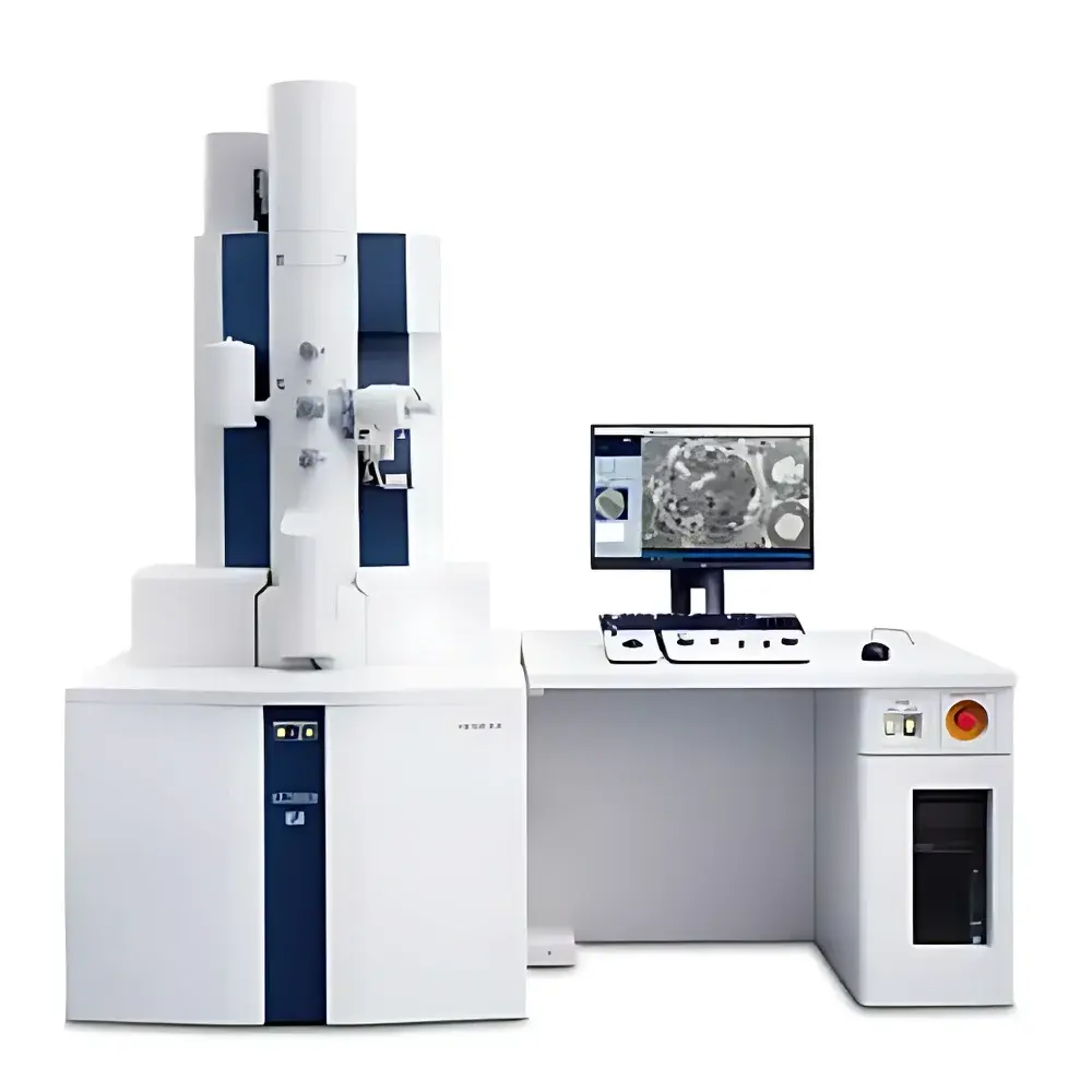
Hitachi HT7800II Transmission Electron Microscope
| Brand | Hitachi |
|---|---|
| Origin | Japan |
| Manufacturer | Hitachi High-Tech Corporation |
| Product Type | Imported TEM |
| Model | HT7800II |
| Accelerating Voltage | 20–120 kV |
| Magnification | ×200–×200,000 (High-Contrast Mode) |
Overview
The Hitachi HT7800II is a high-performance, dual-mode transmission electron microscope engineered for precision structural characterization across materials science, life sciences, and nanotechnology research. Utilizing Hitachi’s proprietary compound objective lens design, the HT7800II uniquely integrates two distinct optical configurations—long-focal-length (high-contrast) and short-focal-length (high-resolution)—within a single objective lens assembly. This architecture eliminates mechanical lens exchange or physical reconfiguration, enabling rapid, reproducible switching between imaging modes without compromising optical alignment or column stability. In high-contrast mode, the instrument delivers exceptional signal-to-noise ratio and wide-field imaging capability ideal for thick biological sections, polymer blends, and hydrated soft matter. In high-resolution mode, it achieves sub-nanometer lattice resolution suitable for atomic-scale analysis of crystalline nanoparticles, graphene, catalytic surfaces, and viral capsids. The system operates over a continuously adjustable accelerating voltage range of 20–120 kV, allowing optimized beam-sample interaction for radiation-sensitive specimens while maintaining sufficient penetration for inorganic thin films.
Key Features
- Dual-mode compound objective lens: Enables seamless, software-controlled transition between high-contrast (HC) and high-resolution (HR) imaging—no manual lens swapping or recalibration required.
- Intuitive GUI-driven operation: All functions—including stage navigation, magnification selection, camera control, focus/stigmation, and image acquisition—are consolidated on a single high-resolution monitor interface, reducing operator training time and procedural variability.
- Standard automatic beam alignment (ABA): A real-time, algorithm-based electron beam centering and stigmation routine that compensates for gun drift, lens hysteresis, and environmental perturbations—ensuring consistent illumination conditions across users and sessions.
- Integrated tomography capability: Built-in tilt-series acquisition support (±70° mechanical tilt range) with synchronized image capture and dose-symmetric data collection protocols for robust 3D reconstruction via weighted back-projection or iterative algorithms.
- Modular expansion architecture: Designed to accommodate optional add-ons including cryo-transfer systems, CLEM correlation stages, STEM detectors, and EDS spectrometers—all interfaced through standardized vacuum and electrical ports compliant with JEOL/Hitachi TEM platform conventions.
Sample Compatibility & Compliance
The HT7800II supports a broad spectrum of specimen types—from conventional epoxy-embedded ultrathin sections (50–100 nm) and carbon-coated grids to plunge-frozen vitreous ice specimens (using optional cryo-stage). Its low-dose imaging protocols, beam blanking synchronization, and variable kV operation meet established criteria for cryo-EM workflow compliance per EMDB and PDB deposition guidelines. When configured with EDS and STEM modules, the system adheres to ASTM E1508–21 for quantitative microanalysis and ISO 16700:2016 for electron probe microanalysis reporting. Full audit trails, user access logs, and parameter versioning are supported when operated under GLP-compliant software environments (e.g., via third-party LIMS integration).
Software & Data Management
The microscope is controlled by Hitachi’s TEM Control Station software, featuring native DICOM-compatible image export, TIFF/DM3 metadata embedding, and direct integration with common reconstruction pipelines (e.g., IMOD, RELION, Dynamo). Optional “EM Flow Creator” enables creation of validated, step-by-step workflows—including automated focus search, multi-region montage acquisition, tilt-series scheduling, and post-acquisition FFT filtering—each assignable to user roles and executable with one-click initiation. All acquired images retain embedded acquisition parameters (kV, magnification, exposure time, spot size, aperture selection), satisfying FDA 21 CFR Part 11 requirements for electronic records when deployed with appropriate validation documentation.
Applications
- Materials characterization: Crystallographic phase identification, dislocation mapping, grain boundary analysis, and in situ heating/cooling experiments (with compatible holders).
- Cryo-structural biology: Single-particle analysis (SPA) of macromolecular complexes, subtomogram averaging of cellular organelles, and correlative light-electron microscopy (CLEM) of fluorescently tagged structures.
- Nanotechnology development: Size distribution quantification of quantum dots, lattice strain measurement in heteroepitaxial films, and defect engineering validation in 2D materials.
- Failure analysis & quality control: Cross-sectional inspection of semiconductor interconnects, delamination assessment in multilayer packaging, and contaminant identification in pharmaceutical excipients.
FAQ
What is the difference between high-contrast and high-resolution modes on the HT7800II?
High-contrast mode uses a longer focal length configuration optimized for depth of field and signal efficiency—ideal for thicker or low-contrast biological specimens. High-resolution mode employs a shorter focal length with minimized spherical aberration for maximum spatial resolution at high magnifications.
Is the HT7800II compatible with automated cryo-EM workflows?
Yes—when equipped with the optional cryo-transfer system and Gatan K2/K3 direct detection camera, the HT7800II supports dose-fractionated movie acquisition, motion correction, and CTF estimation within standard cryo-EM processing stacks.
Can the HT7800II perform elemental mapping?
Elemental analysis is enabled via optional STEM and EDS modules, supporting qualitative and semi-quantitative X-ray mapping with spatial resolution down to ~1 nm under optimal conditions.
Does the system support remote operation and data sharing?
The TEM Control Station includes secure VNC-based remote access, role-based permission controls, and configurable network storage paths compliant with institutional IT security policies.
What maintenance intervals are recommended for long-term operational stability?
Hitachi recommends quarterly vacuum system checks, biannual filament replacement (if using tungsten or LaB₆), and annual column alignment verification—documentation and service logs are automatically archived within the system database.


