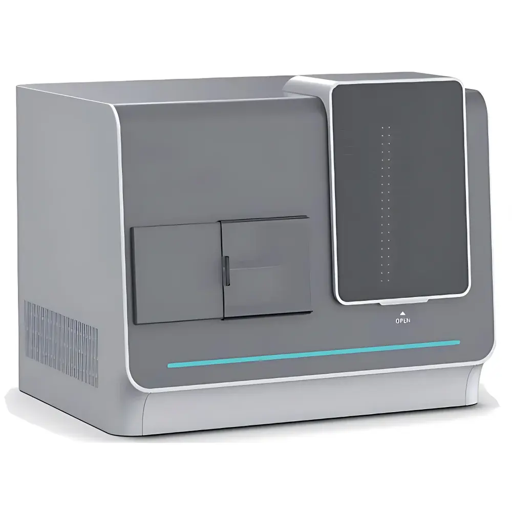
Lei-Tech LK-PCYG Fully Automated Inverted Fluorescence Microscope
| Brand | Lei-Tech |
|---|---|
| Origin | Tianjin, China |
| Manufacturer Type | Direct Manufacturer |
| Product Category | Domestic |
| Model | LK-PCYG |
| Instrument Type | Inverted Fluorescence Microscope |
| Medical Device Classification | Non-Medical Device |
| Instrument Class | Research-Grade Fluorescence Microscope |
| Slide Capacity | 3 / 30 / 60 / 120 slides |
| Scan Speed | ≤50 s @ 20×, <80 s @ 40× (15 mm × 15 mm FOV) |
| Scanning Method | Continuous Area Scan |
| Objective Configuration | Motorized 20× (optional 40× with turret) |
| Stage | Dual-Heterostructure Precision Stage |
| Motion Accuracy | XYZ = 0.1 µm |
| Software Features | Global Mosaic Stitching, Extended Depth-of-Field (EDF) Fusion, Lossless TIFF/SVS Export |
| Preview Functionality | Infinite Zoom, Annotation, Screenshot |
| Power Supply | AC 220 V ±10% |
| Operating Temperature | 5–40 °C |
Overview
The Lei-Tech LK-PCYG Fully Automated Inverted Fluorescence Microscope is a research-grade digital pathology imaging platform engineered for high-fidelity, high-throughput fluorescence and brightfield scanning of biological specimens. Built on an inverted optical architecture, it enables stable, vibration-resistant observation of live or fixed samples in culture vessels—ideal for adherent cell monolayers, tissue sections, cytology smears, and immunohistochemically stained slides. Its core imaging methodology integrates short-exposure high-speed line-scan acquisition with adaptive continuous autofocus, leveraging closed-loop multi-axis synchronization and magnetic-levitation Z-control to maintain sub-micron focus stability across large-area scans. Unlike conventional point-scanning systems, the LK-PCYG employs a dual-heterostructure linear motor stage and low-distortion, large-aperture optics to achieve seamless, distortion-corrected mosaic stitching at 20× and 40× magnifications—ensuring quantitative spatial fidelity required for downstream AI-assisted analysis, telepathology, and longitudinal morphometric studies.
Key Features
- Adaptive real-time autofocus: Magnetic-levitation Z-drive with 0.1 µm resolution ensures consistent focus depth across heterogeneous sample topographies—including uneven tissue sections and thick 3D cultures.
- High-speed continuous area scanning: Achieves full-slide capture in ≤50 seconds at 20× (15 mm × 15 mm field), with optional 40× motorized objective turret for higher-resolution subregion analysis.
- Dual-heterostructure precision stage: Combines orthogonal linear motor axes with sub-micron repeatability (XYZ = 0.1 µm), minimizing mechanical drift during extended acquisition protocols.
- Optimized optical path: Custom-designed low-aberration objectives and high-quantum-efficiency sCMOS detection support multi-channel fluorescence (DAPI/FITC/TRITC/Cy5) with minimal crosstalk and photobleaching.
- Integrated hardware-software co-design: Onboard FPGA-accelerated image processing enables real-time EDF fusion, automatic ROI detection, and lossless TIFF/SVS export compliant with DICOM-SR and ASAM standards.
Sample Compatibility & Compliance
The LK-PCYG accommodates standard 25 mm × 75 mm glass slides, supporting up to 120-slide unattended batch operation via programmable cassette loading. It is validated for formalin-fixed paraffin-embedded (FFPE) tissue sections, liquid-based cytology (LBC) preparations, chromosome spreads, and microarray chips. While not classified as a medical device under FDA 21 CFR Part 809 or EU IVDR, the system meets ISO 13485-aligned manufacturing controls and supports GLP/GMP-compliant workflows through audit-trail-enabled software logging, user-role access control, and electronic signature capability (per FDA 21 CFR Part 11 Annex A). All exported SVS files conform to OpenSlide-compatible pyramidal tiling, enabling interoperability with PathAI, QuPath, HALO, and commercial PACS environments.
Software & Data Management
The proprietary LK-ScanSuite™ software provides a unified interface for acquisition, visualization, annotation, and export. Core modules include: (1) Auto-ROI detection using contrast-adaptive thresholding and morphological segmentation; (2) Global mosaic stitching with intensity normalization and seam blending; (3) Extended depth-of-field synthesis via Z-stack fusion; (4) Real-time annotation overlay with DICOM-SR metadata embedding; and (5) Cloud-sync gateway supporting encrypted HTTPS upload to AWS S3, Azure Blob, or on-premise NAS. All raw data can be archived in uncompressed TIFF or compressed SVS format—preserving pixel-level integrity for retrospective reanalysis. Software updates are delivered via secure OTA channels with SHA-256 verified binaries.
Applications
The LK-PCYG serves as a foundational imaging engine in academic pathology labs, biotech QC facilities, and translational research centers. Primary use cases include: digital slide archiving for longitudinal cohort studies; AI training dataset generation for cervical cytology (e.g., ASC-US triage), nuclear morphology quantification, and mitotic figure detection; automated karyotype analysis via high-resolution chromosome spread scanning; and remote collaborative diagnosis through synchronized multi-user viewing on web-native viewers. Its non-contact, high-speed scanning protocol eliminates manual focusing variability—enhancing inter-laboratory reproducibility in proficiency testing and external quality assessment (EQA) programs accredited by CAP and UK NEQAS.
FAQ
Is the LK-PCYG compliant with FDA 21 CFR Part 11 for electronic records?
Yes—LK-ScanSuite™ includes role-based authentication, audit trails, electronic signatures, and immutable log archiving, meeting all technical requirements for regulated environments.
Can the system integrate with existing LIS/PACS infrastructure?
Yes—it supports HL7 v2.x messaging, DICOM-SR export, and RESTful API endpoints for bidirectional integration with major laboratory information systems.
What is the maximum supported slide thickness for autofocus reliability?
The magnetic-levitation Z-drive maintains stable focus across samples up to 1.2 mm in height, including standard microscope slides with coverslips and thick-sectioned organoids.
Does the system support time-lapse live-cell imaging?
While optimized for static high-resolution scanning, optional environmental chamber integration (37°C/5% CO₂) and LED illumination modulation enable limited kinetic acquisition at defined intervals.
Are software updates provided post-purchase, and for how long?
Lei-Tech offers two years of complimentary firmware and software maintenance, including security patches, feature enhancements, and compatibility updates for new OS versions and cloud platforms.


