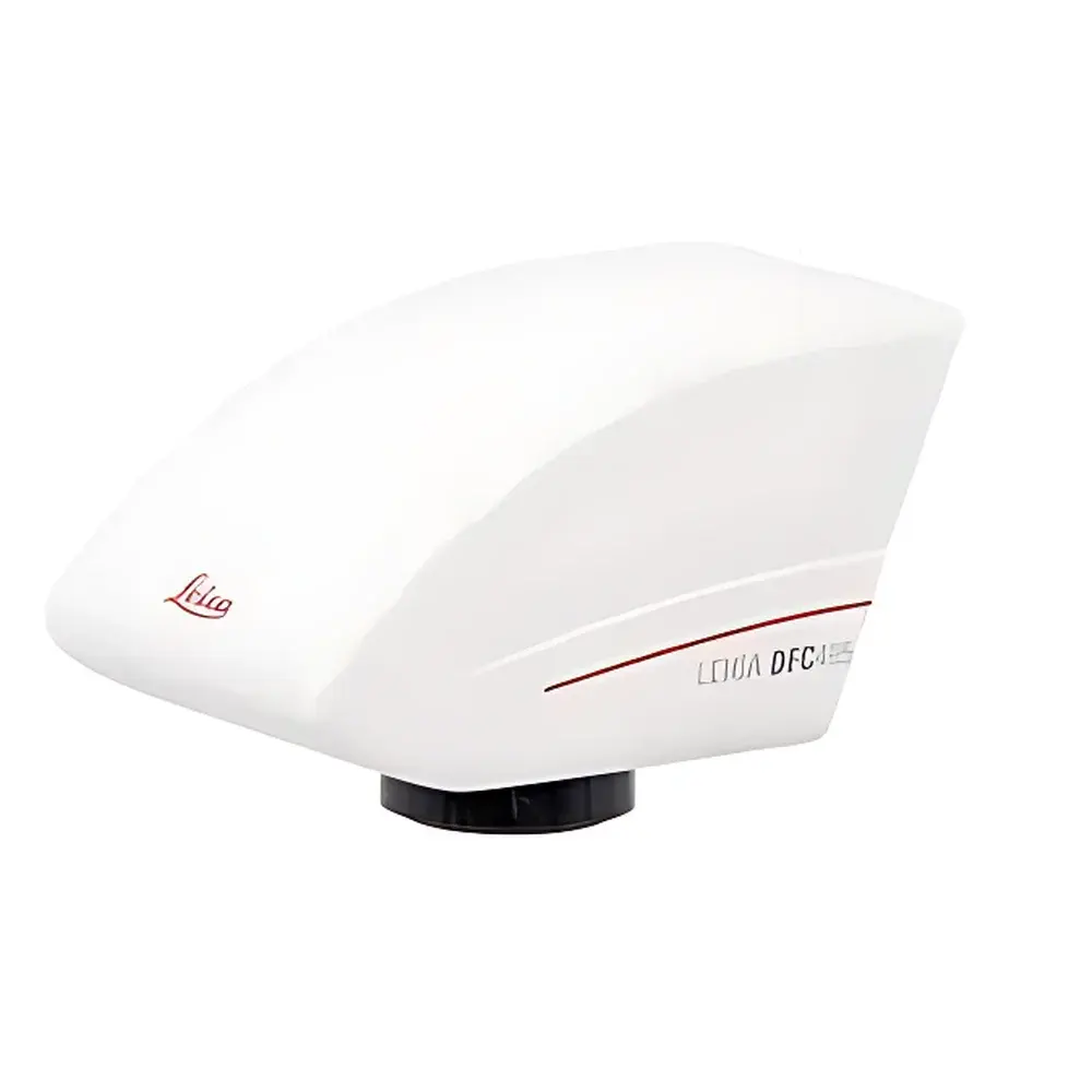
Leica DFC450 Microscope Camera
| Brand | Leica |
|---|---|
| Origin | Germany |
| Model | DFC450 |
| Sensor Type | CCD |
| Resolution | 5 MP (2592 × 1944) |
| Interface | IEEE 1394b (FireWire B) |
| Preview Resolution | SXGA (1280 × 960) @ up to 18 fps |
| Power Delivery | via FireWire bus |
| Software Compatibility | Leica Application Suite (LAS), LAS X, Windows/macOS/Linux (with drivers) |
Overview
The Leica DFC450 is a high-performance, research-grade microscope camera engineered for precision digital imaging in life science, clinical pathology, materials science, and industrial quality control applications. Built upon a progressive-scan CCD sensor architecture, the DFC450 delivers true 5-megapixel resolution (2592 × 1944 pixels) with excellent quantum efficiency and low-noise analog signal processing—enabling faithful color reproduction and high dynamic range across diverse illumination conditions. Its optical design supports C-mount coupling to all major Leica upright, inverted, stereo, and metallurgical microscope platforms—including the DM series, M series, and DMLP/DM750P polarized systems—ensuring seamless integration into existing optical workflows. Unlike CMOS-based alternatives optimized for speed over fidelity, the DFC450 prioritizes photometric accuracy and spatial consistency, making it suitable for quantitative morphometric analysis, archival documentation, and GLP-compliant image acquisition where pixel integrity and repeatability are critical.
Key Features
- 5.0 MP interline-transfer CCD sensor with global shutter operation—eliminates motion artifacts during time-lapse or stage-scanning acquisitions.
- SXGA live preview (1280 × 960) at up to 18 frames per second, enabling real-time focusing, sample positioning, and illumination optimization without latency.
- Integrated FireWire B (IEEE 1394b) interface delivering both data transfer and bus-powered operation—no external power supply required; simplifies cabling and reduces electromagnetic interference in sensitive lab environments.
- Patented sharpness enhancement algorithm applied in-camera, improving edge contrast and microstructural definition without introducing halos or false detail—particularly beneficial for unstained tissue sections, semiconductor surface inspection, and metallographic grain boundary delineation.
- “Save & Recall” function stores complete exposure parameters (gain, exposure time, white balance, gamma, ROI) as named presets—ensuring identical acquisition conditions across longitudinal studies or multi-user labs compliant with ISO/IEC 17025 documentation requirements.
- Full compatibility with Leica’s LAS and LAS X software platforms, supporting automated multi-channel fluorescence capture, Z-stack acquisition, tiling, and metadata embedding per DICOM-SR and MIAME standards.
Sample Compatibility & Compliance
The DFC450 is validated for use across transmitted light (brightfield, phase contrast, DIC), reflected light (epi-fluorescence, darkfield, polarization), and oblique illumination modalities. Its spectral response (400–700 nm) aligns with standard halogen, LED, and mercury/xenon excitation sources used in routine histopathology and semiconductor wafer inspection. The camera meets CE marking requirements under Directive 2014/30/EU (EMC) and 2014/35/EU (LVD), and its firmware implements audit-trail-capable session logging when operated within LAS X in compliance with FDA 21 CFR Part 11 Annex 11 guidelines. It supports acquisition protocols referenced in ASTM E2015 (Standard Guide for Digital Imaging in Microscopy) and ISO 10934-1 (Medical endoscopes — Terminology).
Software & Data Management
Leica Application Suite (LAS) and LAS X provide native driver support for the DFC450, enabling hardware-triggered synchronization with motorized stages, filter wheels, and focus drives. All captured images embed EXIF-compatible metadata—including objective magnification, condenser settings, illumination intensity, and user-defined annotations—facilitating traceability in regulated environments. Raw 16-bit TIFF output preserves linear intensity response for downstream quantification in ImageJ/Fiji, MATLAB, or HALO®. Batch export supports DICOM, OME-TIFF, and JPEG2000 formats with embedded ICC profiles for color-managed display and publication workflows.
Applications
- Quantitative histomorphometry in preclinical toxicology studies (e.g., liver steatosis scoring, renal glomerular area measurement).
- Non-destructive failure analysis of printed circuit boards and MEMS devices using reflected-light brightfield imaging.
- Particle size distribution analysis of airborne contaminants in cleanroom monitoring (per ISO 14644-1 Class 5–8 environments).
- Metallurgical grain structure documentation per ASTM E112 and ISO 643 standards.
- Live-cell imaging support via external TTL triggering for calcium flux assays and mitochondrial dynamics tracking.
- Educational microscopy labs requiring robust, calibration-stable imaging for student annotation and comparative analysis.
FAQ
Is the DFC450 compatible with non-Leica microscopes?
Yes—the C-mount interface and standard FireWire B protocol enable integration with Olympus, Nikon, Zeiss, and custom-built optical systems, provided mechanical back focal distance and flange distance specifications are met.
Does the camera support binning or region-of-interest (ROI) readout?
Yes—hardware-level 2×2 binning is available to improve SNR in low-light fluorescence applications; ROI selection reduces frame transfer time and enables high-speed sub-area monitoring.
Can exposure settings be controlled remotely via API?
Yes—Leica provides a documented COM/ActiveX SDK for Windows and a C++ SDK for Linux/macOS, supporting full parameter automation in custom-developed acquisition software.
What is the maximum cable length supported for FireWire B operation?
Up to 4.5 meters with certified 1394b cables; longer distances require active repeaters or conversion to GigE via compliant bridge devices.
Is firmware update capability included?
Yes—firmware updates are distributed through Leica’s official support portal and applied via LAS X; version history and release notes are publicly archived for audit purposes.


