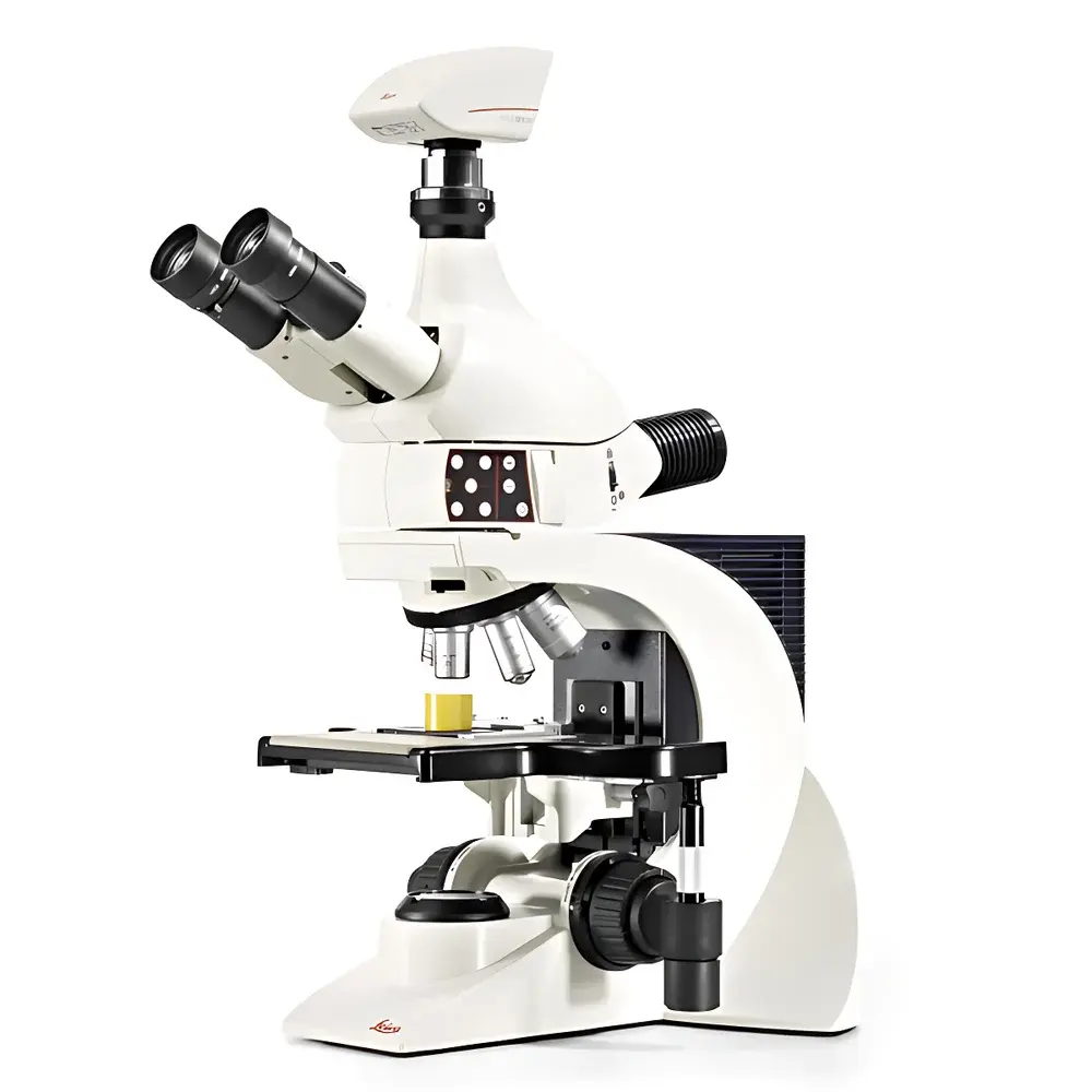
Leica DM1750 M Metallurgical Microscope
| Brand | Leica |
|---|---|
| Origin | Germany |
| Model | DM1750 M |
| Configuration | Upright |
| Total Magnification | 1050× |
| Eyepieces | 5× BF/DF M32 or 6× BF M25 or 7× BF M25 |
| Objective Lenses | Up to 150× |
| Integrated Image Analysis System | Yes |
| Illumination | High-Power White LED (Cold Light, >20,000 h Lifetime) |
| Condenser | Built-in Adjustable Aperture Diaphragm |
| Objective Turret | 6- or 7-Position, Precision-Centered |
| Stage Surface | Ultra-Hard Ceramic Coating |
| Maximum Specimen Height | 80 mm |
| Tilted Illumination | Quad-LED Oblique Lighting Module |
| Optical Path | Optimized Reflected-Light Path for BF, DF, and Polarized Light |
Overview
The Leica DM1750 M is a high-performance upright metallurgical microscope engineered for precision microstructural analysis of opaque, reflective materials—including metals, alloys, ceramics, composites, and coated surfaces. Its optical architecture is based on a fully corrected, infinity-corrected reflected-light path optimized for brightfield (BF), darkfield (DF), and polarized light (POL) observation—enabling rigorous qualitative and quantitative evaluation in industrial QA/QC, failure analysis, and academic metallurgy laboratories. Designed and assembled in Wetzlar, Germany, the DM1750 M integrates robust mechanical stability with modular illumination flexibility, ensuring reproducible imaging performance even under variable ambient conditions typical of production-floor or shared core-facility environments.
Key Features
- Precision-reflected optical path: Engineered for minimal chromatic and spherical aberration across BF, DF, and POL modalities—ensuring accurate grain boundary delineation, phase contrast differentiation, and inclusion identification.
- High-efficiency white LED illumination: Cold-white, 5700 K LED source with >20,000 hours rated lifetime; integrated adjustable aperture diaphragm enables precise control of numerical aperture and contrast without thermal drift or filament degradation.
- Quad-LED oblique illumination system: Four independently controllable LEDs mounted at fixed asymmetric angles provide directional contrast enhancement—critical for detecting micro-scratches, surface topography variations, and subsurface defects without mechanical stage tilting.
- 6- or 7-position precision objective turret: CNC-machined, kinematically aligned turret guarantees parfocality and centration across all objectives (including up to 150×), eliminating refocusing and recentering during magnification changes.
- Ultra-hard ceramic stage surface: Chemically inert, scratch-resistant alumina-toughened ceramic coating withstands repeated abrasion from coarse metallographic samples and heavy instrumentation—maintaining flatness and positional accuracy over years of high-throughput use.
- Large specimen capacity: Accommodates specimens up to 80 mm in height and standard 76 × 52 mm slide dimensions—compatible with automated sample holders and motorized Z-stage upgrades for serial sectioning workflows.
Sample Compatibility & Compliance
The DM1750 M supports standardized metallurgical sample preparation per ASTM E3, ISO 643, and EN 10360. Its rigid base and vibration-damped column ensure stability during long-duration imaging and microhardness correlation studies. The microscope complies with IEC 61000-6-3 (EMC emission standards) and IEC 61000-6-2 (immunity), meeting requirements for integration into GLP- and GMP-regulated environments. Optional traceable calibration kits (NIST-traceable stage micrometers and resolution test targets) support ISO/IEC 17025-compliant instrument qualification protocols.
Software & Data Management
When paired with Leica Application Suite X (LAS X) software, the DM1750 M delivers full digital workflow integration—including real-time image acquisition, multi-channel overlay (BF/DF/POL), particle analysis (grain size per ASTM E112), phase fraction quantification, and automated report generation. LAS X supports FDA 21 CFR Part 11 compliance via electronic signatures, audit trails, and role-based user access control—enabling secure data handling in regulated manufacturing and clinical research settings. Export formats include TIFF (16-bit), JPEG2000, and CSV for statistical post-processing in MATLAB or JMP.
Applications
- Microstructural characterization of heat-treated steels, aluminum alloys, and nickel-based superalloys
- Failure analysis of fracture surfaces, fatigue cracks, and intergranular corrosion
- Coating thickness measurement and adhesion defect mapping
- Quality control of sintered powders, additive-manufactured components, and brazed joints
- Research-grade investigations in physical metallurgy, phase transformation kinetics, and recrystallization behavior
- Education and training in materials science curricula requiring ISO-standardized imaging practices
FAQ
Is the DM1750 M compatible with third-party camera systems?
Yes—the microscope features a standardized C-mount port (1× or 0.63× reduction optics) and TTL synchronization interface, supporting most scientific CMOS/CCD cameras with GenICam or USB3 Vision drivers.
Can the quad-LED oblique lighting be used simultaneously with polarized light?
Yes—polarizer and analyzer modules are mounted in the optical path upstream of the illumination collector, allowing independent activation of oblique contrast and polarization states.
What maintenance is required for the ceramic stage surface?
No routine polishing or recoating is necessary; cleaning requires only lint-free wipes and isopropyl alcohol—no abrasive cleaners or solvents are recommended.
Does the DM1750 M support motorized focus or stage automation?
Motorized Z-focus and XY stage options are available as factory-installed accessories and integrate natively with LAS X for scripting and macro-based batch acquisition.
How is calibration verified for quantitative measurements?
Calibration is performed using certified stage micrometers and resolution test targets traceable to NIST SRM 2059 and ISO 10962; verification reports are generated within LAS X’s instrument qualification module.


