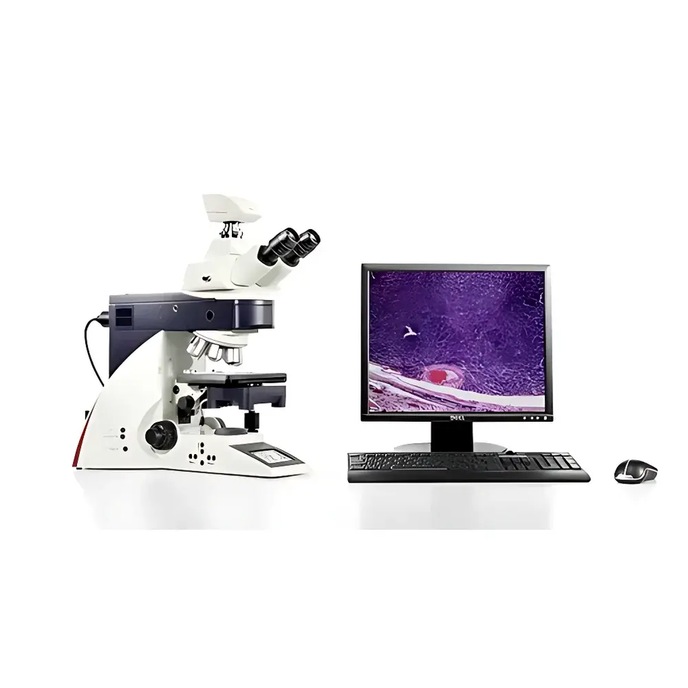
Leica DM4000B Biological Microscope
| Brand | Leica |
|---|---|
| Origin | Germany |
| Model | DM4000B |
| Instrument Type | Upright Biological Microscope |
| Illumination | LED Transmitted Light (12 V, 100 W Halogen Optional) |
| Optical Path | Infinity-Corrected |
| Objective Turret | 6-Position Encoded Motorized |
| Fluorescence Capability | Motorized 5-Position Filter Cube Holder with Leica Zero-Drift Technology |
| Contrast Methods | Brightfield, Phase Contrast, Darkfield, Polarization, DIC (Automated) |
| Eyepiece Field of View | 25 mm |
| Observation Tube | Binocular or Trinocular |
| Software Integration | Leica Application Suite (LAS), LAS AF for Advanced Fluorescence Imaging |
| Compliance | Designed for GLP/GMP environments |
Overview
The Leica DM4000B Biological Microscope is an upright, research-grade optical platform engineered for precision life science applications—from routine histopathology to advanced live-cell fluorescence imaging. Built on Leica’s proprietary infinity-corrected optical architecture, the DM4000B integrates a fully encoded, motorized 6-position objective turret with intelligent illumination management to deliver consistent, high-fidelity image acquisition across modalities including brightfield, phase contrast, darkfield, polarization, and differential interference contrast (DIC). Its core design principle centers on operational reproducibility: automated Köhler illumination alignment, objective-synchronized light intensity adjustment, and real-time parameter feedback via integrated OLED display ensure minimal user-induced variability—critical for longitudinal studies and multi-user laboratory environments.
Key Features
- Motorized, fully encoded 6-position objective turret with dry and oil-immersion compatibility (M25 thread standard)
- LED-transmitted illumination system delivering stable color temperature (5700 K) across full intensity range; rated lifetime ≥50,000 hours
- Optional 12 V / 100 W halogen illumination module for legacy protocol compatibility
- Motorized 5-position fluorescence filter cube holder with Leica Zero-Drift positioning technology for precise multi-channel registration
- Fluorescence Intensity Management (FIM): patented algorithm enabling channel-specific light dose control to minimize photobleaching and ensure quantitative repeatability
- SmartTouch interface with 6 programmable function keys located ergonomically on focus knobs for hands-free operation
- Integrated electronic control unit (CTR5000) housing power supply, motor drivers, and illumination management logic—no external electronics rack required
- Motorized XY stage with position memory (up to 6 locations) and torque-adjustable ceramic-coated mechanical drive
- Automated condenser control: motorized top lens, 7-position aperture disc, and optional motorized polarizer
Sample Compatibility & Compliance
The DM4000B accommodates standard 1″ × 3″ glass slides, petri dishes (up to 100 mm diameter), and multi-well plates (6–96 well) using optional stage adapters. Its modular contrast optics support unstained live specimens (phase contrast), fixed tissue sections (H&E, IHC), birefringent structures (collagen, starch), and fluorescently labeled subcellular targets (GFP, DAPI, TRITC). The system meets essential requirements for regulated environments: when operated with Leica Application Suite Advanced Fluorescence (LAS AF) v4.0+, it supports electronic signatures, role-based user authentication, and full audit trail generation compliant with FDA 21 CFR Part 11. All optical components adhere to ISO 10934-1 (microscope nomenclature) and DIN EN 61000-6-3 (EMC emissions) standards.
Software & Data Management
The DM4000B functions as a hardware node within Leica’s unified imaging ecosystem. Native integration with Leica Application Suite (LAS) enables synchronized control of illumination, focus, stage, and camera—eliminating manual synchronization errors. LAS AF extends functionality to time-lapse multi-dimensional acquisition (x-y-z-t-λ), deconvolution, and co-localization analysis. Image metadata—including objective magnification, NA, illumination intensity, exposure time, and filter configuration—is embedded directly into TIFF/OME-TIFF files. For enterprise deployment, LAS supports DICOM-SR export and PACS integration via HL7/FHIR-compliant gateways. All configuration presets—including user-defined contrast workflows—are stored locally on the microscope’s internal flash memory and can be exported/imported as encrypted XML profiles.
Applications
- Histopathology: Routine H&E and immunohistochemical slide screening with rapid contrast switching and ergonomic repositioning for multi-slide review
- Cell Biology: Long-term live-cell imaging using low-heat LED illumination and FIM-controlled excitation dosing to preserve viability
- Neuroscience: High-contrast phase contrast visualization of primary neuronal cultures and organotypic brain slices
- Microbiology: Differential staining (Gram, acid-fast) with automated brightfield/darkfield transitions and digital annotation
- Materials Life Science Interface: Polarized light analysis of biomineralized tissues (bone, dentin) and synthetic biomaterials
- Quality Control: GMP-compliant documentation of cell morphology, confluence, and contamination status in bioproduction workflows
FAQ
Does the DM4000B support DIC with automatic alignment?
Yes—the system includes motorized Nomarski prism translation and condenser centering, enabling push-button DIC optimization synchronized with objective selection.
Can I retrofit my existing DM4000B with LED illumination?
LED upgrade kits (part no. 11506008) are available for all DM4000B units manufactured after 2012; installation requires factory-certified service technician calibration.
Is LAS AF software included with the microscope purchase?
LAS AF is licensed separately; base LAS is bundled. A perpetual license with annual maintenance (including updates and remote support) is recommended for regulated use.
What is the maximum working distance supported for long-working-distance objectives?
The stage clearance allows up to 25 mm vertical space between objective front lens and specimen plane—compatible with 20×/0.40 WD 6.9 mm and 40×/0.60 WD 3.5 mm objectives.
How does the Zero-Drift fluorescence filter technology improve multi-channel imaging?
It eliminates mechanical hysteresis during filter wheel rotation by using closed-loop stepper motor control and optical position feedback—ensuring sub-micron spatial registration stability across sequential channel acquisitions.


