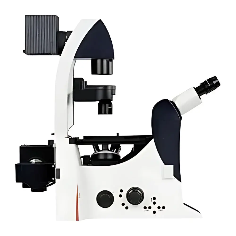
Leica DMI4000 B Automated Inverted Microscope for Life Science Research
| Brand | Leica |
|---|---|
| Origin | Germany |
| Model | DMI4000 B |
| Type | Automated Inverted Biological Microscope |
| Optical Configuration | Fluorescence-Optimized with Motorized Filter Turret and FIM (Fluorescence Intensity Manager) |
| Illumination Management | Motorized Brightfield/DIC/Phase Contrast Switching |
| Condenser | Universal Multi-Technique Condenser (Brightfield, Darkfield, Phase Contrast, DIC, Hoffman Modulation Contrast) |
| Response Time for Filter & Intensity Change | ≤20 ms |
| Fluorescence Intensity Control | 5-step discrete intensity levels with per-filter-block memory |
| Automation Level | Fully motorized objective nosepiece, filter turret, focus, condenser alignment, and illumination aperture control |
Overview
The Leica DMI4000 B is a high-performance automated inverted microscope engineered specifically for demanding life science applications—including live-cell imaging, long-term time-lapse studies, and multi-modal fluorescence analysis in adherent and suspension cultures. Its optical architecture is built around Leica’s proprietary HCX PL FLUOTAR objective series and a fluorescence-dedicated light path featuring an integrated dichroic beam splitter and high-transmission emission filters. The system operates on the principle of transmitted-light contrast enhancement combined with epifluorescence excitation, enabling simultaneous or sequential acquisition across multiple contrast modes without mechanical realignment. Designed for integration into regulated laboratory environments, the DMI4000 B supports GLP-compliant documentation workflows and meets essential optical safety standards (IEC 61000-6-3, EN 61326-1) for laboratory instrumentation.
Key Features
- Fully motorized inverted platform with encoded objective revolver (up to 6 positions), enabling precise, repeatable magnification switching and software-triggered objective recall.
- Integrated Fluorescence Intensity Manager (FIM): electronically regulates LED or metal-halide lamp output across five discrete intensity levels; stores individual intensity settings per fluorescence filter cube to maintain photometric consistency during multi-channel acquisitions.
- High-speed motorized filter turret with ≤20 ms filter exchange—critical for minimizing phototoxicity in live-cell experiments and enabling rapid spectral multiplexing (e.g., GFP/mCherry dual-channel time-lapse).
- Universal multi-contrast condenser (Leica NCG, Numerical Aperture 0.9) supporting brightfield, darkfield, phase contrast (Ph1–Ph4), differential interference contrast (DIC), and Hoffman modulation contrast—all accessible via software-controlled lever actuation or automated preset recall.
- Motorized Köhler illumination alignment system: automatically adjusts field and aperture diaphragms based on selected objective and contrast mode, ensuring optimal resolution, contrast, and signal-to-noise ratio without manual intervention.
- Ergonomic stage design with motorized X-Y translation (130 × 85 mm travel), compatible with environmental chambers (CO₂, temperature, humidity) and multi-well plate holders for high-content screening workflows.
Sample Compatibility & Compliance
The DMI4000 B accommodates standard tissue culture formats including 35 mm–150 mm Petri dishes, chambered coverglasses, multi-well plates (6–384-well), and custom microfluidic devices. Its long-working-distance objectives (e.g., 20×/0.40, 40×/0.60) enable imaging through up to 1.5 mm of culture medium and vessel bottom thicknesses compliant with ANSI/SLAS standards. The system conforms to ISO 13485:2016 (when deployed in IVD-related research), supports audit trails required under FDA 21 CFR Part 11 when used with Leica Application Suite X (LAS X) software, and complies with EU Directive 2014/30/EU (EMC) and 2014/35/EU (LVD). All optical components are certified free of hazardous substances per RoHS 2011/65/EU.
Software & Data Management
Controlled exclusively via Leica Application Suite X (LAS X), the DMI4000 B supports structured experimental metadata capture—including objective ID, filter set, exposure time, Z-stack parameters, and environmental sensor inputs (if integrated). LAS X provides full GLP/GMP traceability: electronic signatures, user access tiers, change logs, and immutable raw data export in TIFF, OME-TIFF, or HDF5 formats. Batch acquisition protocols can be saved, versioned, and deployed across instrument networks. Optional LAS X Live Data module enables real-time quantitative analysis (e.g., fluorescence intensity kinetics, cell count tracking, morphometric profiling) with export to CSV or direct integration into MATLAB or Python-based analysis pipelines.
Applications
- Long-term live-cell imaging of stem cell differentiation, neuronal network development, and cancer cell migration under physiological conditions.
- Multi-parameter fluorescence co-localization studies using ≥4-color labeling with automated Z-stack acquisition and deconvolution-ready image stacks.
- High-content phenotypic screening in 96- and 384-well formats, leveraging automated stage positioning and protocol-driven image capture.
- Quantitative phase imaging of unstained cells (e.g., red blood cell volume estimation, organelle dynamics) using phase contrast or DIC with calibrated pixel-to-micron mapping.
- Time-resolved assessment of calcium flux, mitochondrial membrane potential, or pH changes using ratiometric fluorescent probes and FIM-enabled intensity stabilization.
FAQ
Is the DMI4000 B compatible with third-party cameras and light sources?
Yes—the system features standardized C-mount and M42 interfaces for camera integration, and its lamp housing supports interchangeable metal-halide, LED, and laser launch modules via Leica’s modular illumination platform.
Does it support automated focus maintenance during time-lapse experiments?
Yes—optional hardware-based focus drift compensation (Leica Focus Finder) or software-based autofocus (LAS X AutoFocus) can be implemented, both compatible with environmental chamber operation.
Can acquisition protocols be exported and shared across multiple DMI4000 B systems?
Yes—LAS X protocol files (.lpx) contain all instrument configuration, acquisition parameters, and metadata schemas, enabling cross-platform reproducibility and method transfer validation.
What level of service and calibration support is available outside the EU?
Leica Microsystems maintains certified service centers in North America, APAC, and EMEA; annual performance verification (per ISO/IEC 17025) and optical calibration services are available globally through authorized partners.
How does the system handle photobleaching mitigation during extended fluorescence imaging?
Beyond FIM-based intensity reduction, LAS X includes adaptive exposure control, region-of-interest (ROI)-based illumination gating, and hardware-synchronized shutter timing to minimize cumulative photon dose while preserving temporal fidelity.
Related Products


