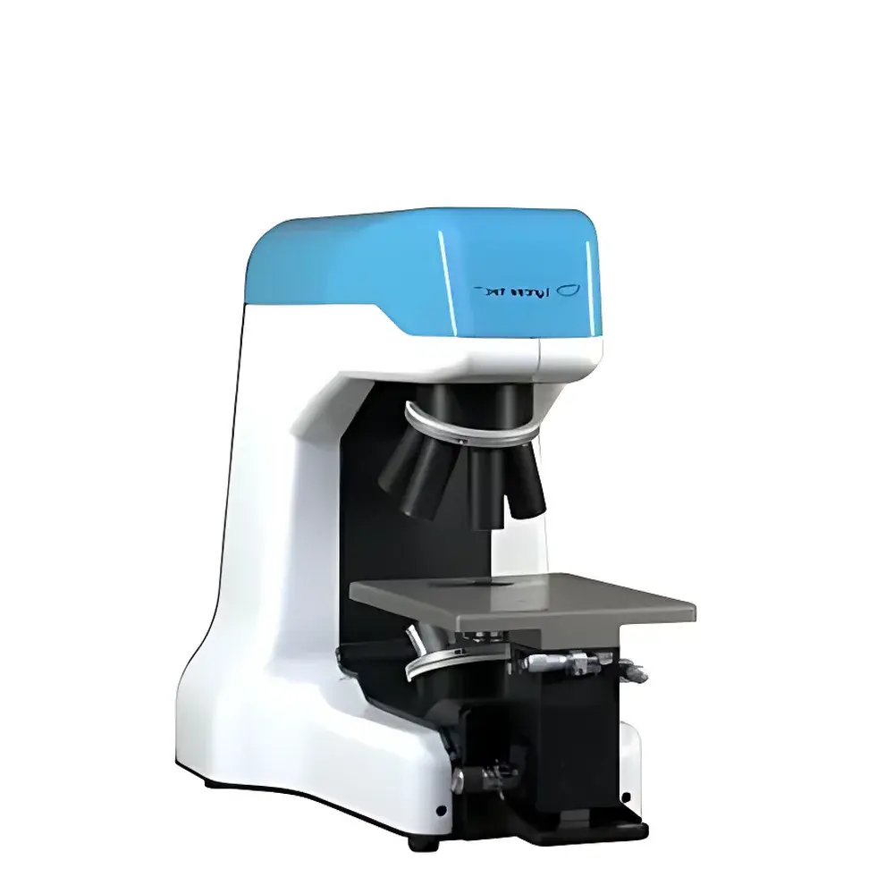
Lyncée Tec DHM-T1000 Transmission Digital Holographic Microscope
| Brand | Lyncée Tec |
|---|---|
| Origin | Switzerland |
| Model | DHM-T1000 |
| Laser Wavelength | 666 nm |
| Vertical Resolution | 2.0 nm |
| Vertical Accuracy | 1.0 nm |
| Repeatability | 0.02 nm |
| Vertical Measurement Range | up to 500 µm |
| Maximum Step Height (Sharp Edge) | up to 1.0 µm |
| Response Time | 500 µs (standard), optional 10 µs |
| Frame Rate | 30 fps (standard), optional up to 1000 fps |
| Hologram Reconstruction Rate | 25 fps (1024×1024 pixels), optional up to 60 fps |
| Lateral Resolution | down to 300 nm (objectives-dependent) |
| Field of View | 66 µm × 66 µm to 5 mm × 5 mm |
| Working Distance | 0.3–18 mm (objectives-dependent) |
| Digital Focus Depth Enhancement | up to 50× depth of field |
| Sample Illumination Intensity | as low as 1 µW/cm² |
Overview
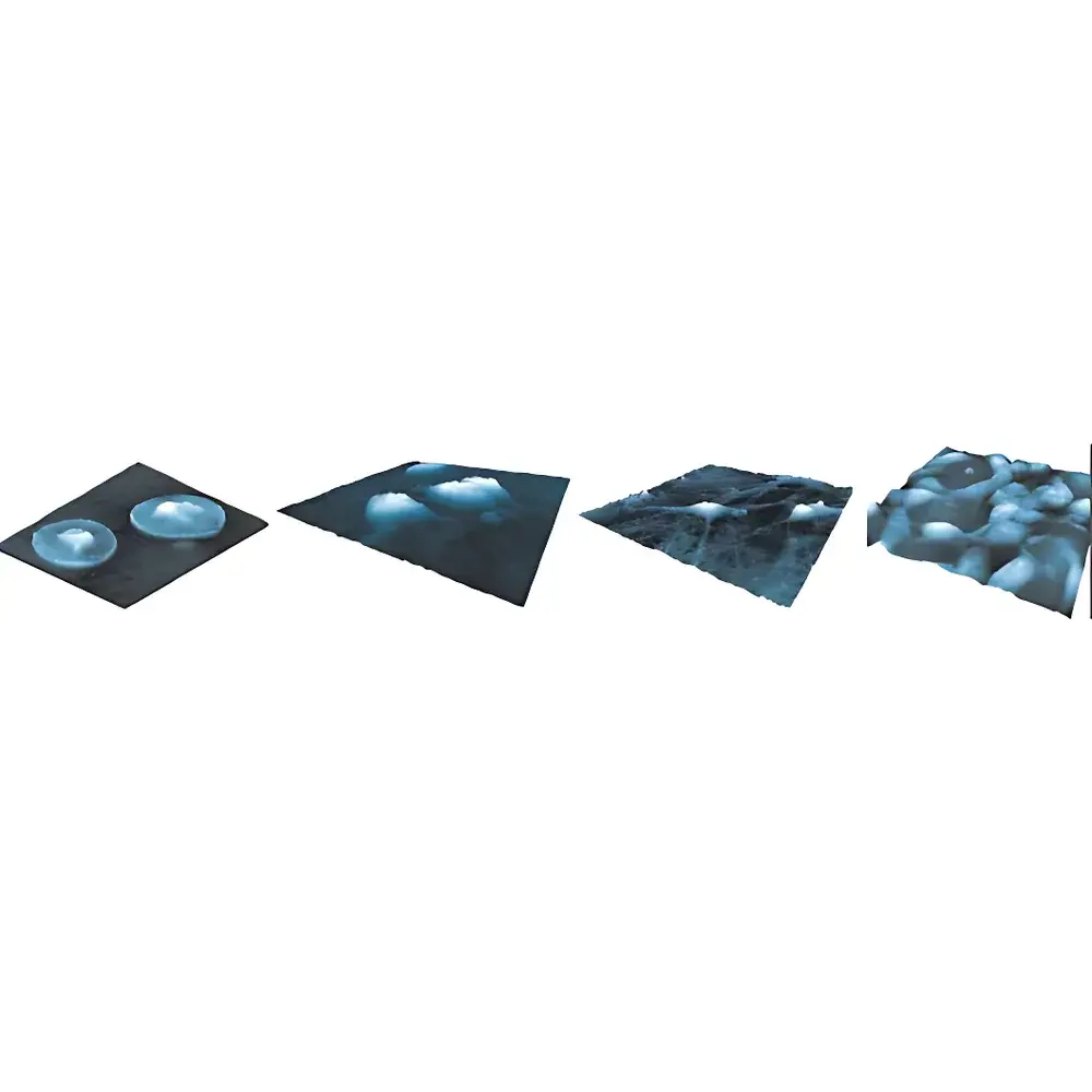
The Lyncée Tec DHM-T1000 is a high-precision, single-wavelength transmission digital holographic microscope engineered for label-free, quantitative phase imaging of transparent and semi-transparent specimens. Unlike conventional brightfield or fluorescence microscopy, the DHM-T1000 operates on the principle of off-axis digital holography: a coherent 666 nm laser beam is split into object and reference arms; interference between the transmitted wavefront through the sample and the undisturbed reference beam generates a digital hologram captured by a high-speed CMOS sensor. This hologram is numerically reconstructed in real time using Fourier-based algorithms to yield both amplitude and quantitative phase maps with nanometric vertical resolution. The system is inherently non-invasive—requiring no staining, fixation, or physical contact—and delivers quantitative optical path difference (OPD) data directly correlated to cellular dry mass, membrane dynamics, refractive index gradients, and subcellular structural changes. Its design targets rigorous applications in live-cell biophysics, micro-optics metrology, and dynamic thin-film characterization where temporal stability, repeatability, and traceable dimensional metrology are essential.
Key Features
- Single-wavelength (666 nm) transmission configuration optimized for high-contrast, low-noise phase imaging of live cells, polymer films, glass substrates, and microfluidic devices
- Nanometric vertical accuracy (1.0 nm) and repeatability (0.02 nm) certified via interferometric calibration using temperature-stabilized narrowband filters (±0.1 nm uncertainty)
- Real-time hologram acquisition at 30 fps standard, extendable to 1000 fps for transient phenomena such as capillary filling, droplet coalescence, or mechanical actuation
- Digital focus synthesis enabling extended depth-of-field imaging—up to 50× greater than conventional microscopy—without mechanical Z-scanning
- Ultra-low phototoxic illumination (<1 µW/cm²), compatible with long-term time-lapse studies of sensitive primary neurons or stem cell monolayers
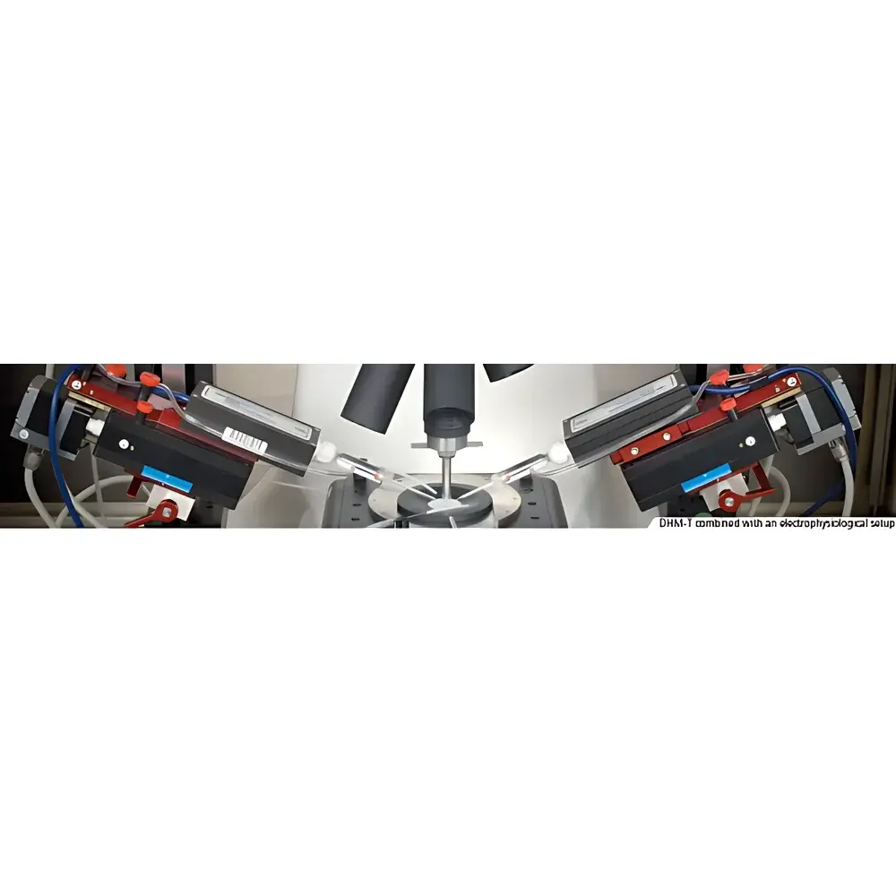
- Modular XYZ stage (114 × 76 × 38 mm travel range) supporting manual or motorized positioning; compatible with environmental chambers and perfusion systems
- Interchangeable objective turret (6-position) accommodating 1.25× to 100× magnifications, including high-NA, long-working-distance, and water/oil-immersion optics
Sample Compatibility & Compliance
The DHM-T1000 is validated for quantitative measurement of optically transmissive samples ranging from submicron-thick lipid bilayers to 500 µm-thick hydrogels and fused silica wafers. It meets foundational requirements for GLP-compliant workflows: raw holograms and reconstructed phase maps are stored with embedded metadata (timestamp, laser power, objective ID, calibration state), supporting audit trails per FDA 21 CFR Part 11 when integrated with compliant LIMS environments. While not a medical device, its measurement traceability aligns with ISO/IEC 17025 principles for dimensional metrology laboratories. Applications include ASTM E2941-22 (optical thickness of transparent coatings) and ISO 25178-603 (areal surface texture analysis via phase-shifting interferometry equivalents). No sample preparation—no dehydration, no conductive coating—is required for static or dynamic topographic profiling.
Software & Data Management
Koala® software—a proprietary C++/C# platform built on .NET Framework—provides full control over acquisition, reconstruction, and analysis. Core modules include real-time phase unwrapping, optical thickness mapping, refractive index inversion (given known substrate geometry), and multi-frame temporal registration. Optional add-ons support automated well-plate scanning (for 96-/384-well assays), particle tracking velocimetry (PTV), and correlation-based strain field analysis. All processed data export in HDF5, TIFF (with embedded OME metadata), or CSV formats; raw holograms are saved losslessly in binary format. Software architecture supports deterministic reconstruction pipelines—enabling reproducible batch processing across instruments—and integrates with Python via documented REST API for custom algorithm deployment in QC/QA validation protocols.
Applications
- Label-free longitudinal monitoring of single-cell proliferation, apoptosis, and drug-induced morphological shifts in adherent and suspension cultures
- Quantitative thickness and uniformity mapping of spin-coated photoresists, anti-reflective coatings, and OLED stack layers
- In situ measurement of swelling kinetics in hydrogels and dissolution profiles of pharmaceutical films
- Dynamic characterization of liquid crystal reorientation under electric fields (response times <10 ms resolvable)
- Defect detection and internal stress analysis in bonded glass substrates and micro-optical assemblies
- High-speed interferometric flow visualization in microchannels—capturing laminar-to-turbulent transition and meniscus dynamics
- Correlative multimodal imaging when paired with optional fluorescence modules, minimizing photobleaching while preserving spatial registration
FAQ
Is the DHM-T1000 suitable for thick, scattering biological tissues?
No—the transmission configuration requires optical transparency; samples exceeding ~100 µm in thickness or exhibiting strong Mie scattering (e.g., dense collagen gels) degrade hologram contrast and reconstruction fidelity.
Can phase data be converted to absolute dry mass values?
Yes—using the established conversion factor of 0.18 mL/g for protein-rich cytoplasm, quantitative dry mass per pixel is derivable from optical path difference maps, validated against coulter counter and QCM-D benchmarks.
Does the system support automated focus maintenance during long-term experiments?
Yes—Koala software includes a closed-loop autofocus routine based on sharpness maximization of reconstructed amplitude images, configurable for periodic or event-triggered correction.
What calibration standards are supplied with the instrument?
A NIST-traceable step-height standard (SiO₂ on Si, nominal 100 nm and 500 nm steps) and interferometric wavelength reference filter are included for initial setup and periodic verification.
Is remote operation supported for facility-shared instruments?
Yes—Koala supports secure remote desktop access and headless acquisition via SSH-triggered scripts; all reconstruction and analysis can be offloaded to local workstations or centralized GPU servers.


