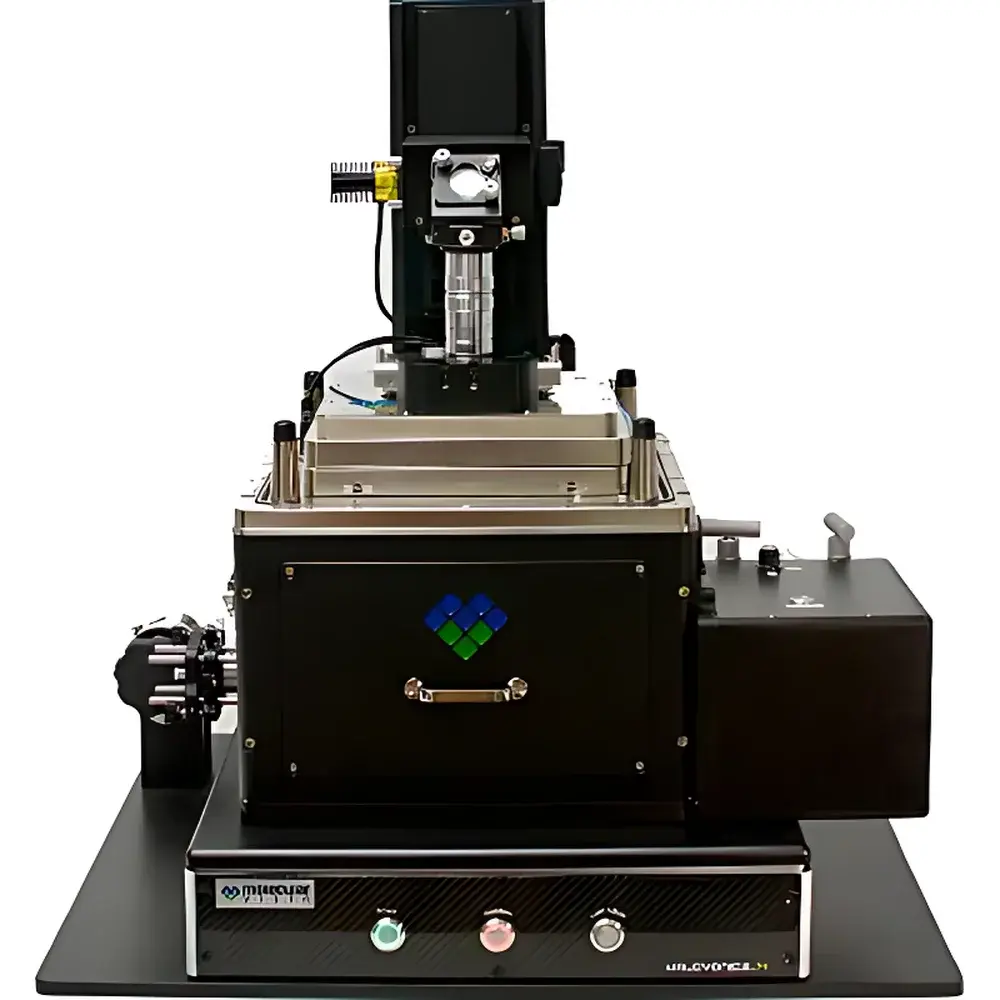
MVI Vista-IR Nanoscale Infrared Imaging and Spectroscopy System
| Brand | MVI |
|---|---|
| Origin | USA |
| Manufacturer Type | Authorized Distributor |
| Product Origin | Imported |
| Model | Vista-IR |
| Instrument Category | Laser-Based Infrared Spectrometer |
| Instrument Type | Laboratory Benchtop System |
| Wavenumber Range | 760–4400 cm⁻¹ |
| Spatial Resolution | <10 nm |
| Spectral Resolution | <2 cm⁻¹ |
| Scan Rate | 10 spectra/sec |
| Signal-to-Noise Ratio | 100:1 |
Overview
The MVI Vista-IR Nanoscale Infrared Imaging and Spectroscopy System is a laboratory-grade, AFM-coupled infrared chemical imaging platform engineered for sub-diffraction-limit spatial resolution. Unlike conventional Fourier Transform Infrared (FTIR) spectrometers—whose lateral resolution is fundamentally constrained by the optical diffraction limit (~3–10 µm at mid-IR wavelengths)—the Vista-IR leverages photo-induced force microscopy (PiFM), a near-field technique that transduces infrared absorption into nanomechanical forces detected by a sharp atomic force microscope (AFM) probe. This eliminates reliance on far-field optics, interferometers, or thermal expansion-based detection mechanisms. By integrating a tunable quantum cascade laser (QCL) source with a high-stability AFM base, the system delivers true nanoscale vibrational contrast with spectral fidelity matching standard FTIR reference libraries. It operates across the functional-group-sensitive mid-IR range (760–4400 cm⁻¹), enabling label-free, chemically specific mapping of organic polymers, inorganic frameworks, catalytic surfaces, and heterogeneous thin films at <10 nm spatial resolution—orders of magnitude beyond conventional IR microscopy.
Key Features
- Sub-10 nm spatial resolution enabled by PiFM detection—immune to lateral thermal expansion artifacts common in photothermal AFM-IR methods
- High-fidelity spectral acquisition with <2 cm⁻¹ spectral resolution, fully compatible with commercial FTIR spectral databases (e.g., SDBS, Hummel, Polymer Library)
- Real-time spectral acquisition at up to 10 spectra per second, supporting dynamic mapping and rapid compositional screening
- Integrated QCL-based tunable IR source delivering high brightness and narrow linewidth across the full 760–4400 cm⁻¹ range
- Benchtop laboratory configuration with vibration-isolated optical table integration and environmental enclosure options for humidity/CO₂ control
- Quantitative amplitude and phase channel output for dual-parameter chemical contrast generation
Sample Compatibility & Compliance
The Vista-IR accommodates a broad range of solid-state samples including polymer thin films (e.g., PES, PS-PMMA block copolymers), catalytic zeolites (e.g., ZSM-5 with variable Si/Al ratios), battery electrode cross-sections, semiconductor heterostructures, and biological membranes. No conductive coating or vacuum requirement is necessary; ambient or controlled-atmosphere operation is standard. The system supports ASTM E1252-98 (Standard Practice for General Techniques for Infrared Microanalysis) and aligns with ISO 1833-10:2021 (Textiles — Quantitative chemical analysis — Part 10: Mixtures of cellulose and polyester) for micro-domain identification. Data acquisition workflows are designed to meet GLP and GMP documentation requirements, with optional audit-trail-enabled software modules compliant with FDA 21 CFR Part 11 for regulated environments.
Software & Data Management
Vista-IR is operated via proprietary MVI Control Suite v4.x—a modular, scriptable platform supporting batch spectral acquisition, hyperspectral cube generation, multivariate curve resolution (MCR), and peak-fitting with Voigt deconvolution. All raw interferograms (where applicable), PiFM amplitude/phase signals, topographic AFM data, and metadata (laser wavelength, dwell time, gain settings, environmental conditions) are stored in vendor-neutral HDF5 format. Integrated spectral library search includes correlation matching against >25,000 reference spectra, with customizable thresholding and false-discovery rate control. Export options include ASCII, CSV, and MATLAB-compatible .mat files; third-party interoperability is supported via Python API (PyVistaIR) for custom machine learning pipelines.
Applications
- Nanoscale phase segregation analysis in multi-component polymer blends and block copolymers
- In situ monitoring of coke deposition and active-site evolution in zeolite catalysts during methanol-to-hydrocarbons (MTH) reactions
- Chemical mapping of interfacial degradation layers in solid-state battery electrodes
- Identification and distribution quantification of crystalline vs. amorphous domains in pharmaceutical co-crystals
- Vibrational fingerprinting of 2D materials (e.g., h-BN, MoS₂ edge states) and their heterostructure interfaces
- Correlative nanoscale IR + Raman + conductive-AFM studies on organic electronic thin films
FAQ
How does PiFM differ from traditional AFM-IR or s-SNOM?
PiFM detects the gradient of the optically induced dipole–dipole interaction between the IR-illuminated sample and conductive AFM tip, rather than photothermal expansion or scattered near-field fields—yielding higher signal-to-noise and eliminating thermal diffusion artifacts.
Can Vista-IR data be directly compared to benchtop FTIR spectra?
Yes—the PiFM-derived absorbance-like spectra exhibit peak positions, relative intensities, and line shapes consistent with transmission/reflection FTIR, enabling direct database matching without empirical calibration transfer.
Is vacuum operation required?
No—ambient air operation is standard; optional purge gas (dry N₂ or Ar) or environmental chamber integration is available for moisture-sensitive samples.
What sample preparation is needed?
Minimal: flat, conductive or semi-conductive substrates (e.g., Si wafers, ITO glass, Au-coated mica); no metal coating required for insulating samples due to non-contact PiFM force detection.
Does the system support time-resolved measurements?
Yes—pulsed QCL operation enables pump-probe mode with temporal resolution down to ~10 ns, suitable for studying ultrafast vibrational relaxation in photoactive materials.


")