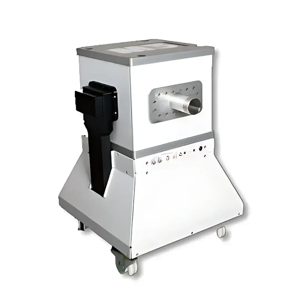
NIUMAG Aspect M3 Small Animal MRI System
| Brand | NIUMAG |
|---|---|
| Origin | Israel |
| Manufacturer Type | Authorized Distributor |
| Origin Category | Imported |
| Model | M3 |
| Instrument Type | Preclinical Magnetic Resonance Imaging (MRI) System |
| Magnet Type | Permanent Magnet, 1.0 T |
| Spatial Resolution | ≤ 235 µm (isotropic) |
| Scan Time | As low as 8–9 min per sequence |
| Cooling | Passive (No Cryogens or Chiller Required) |
| Shielding | Self-Shielded, Zero Stray Field |
| Regulatory Compliance | CE Marked, Designed for GLP-Compliant Preclinical Research |
| Application Focus | Mouse Phenotyping, Longitudinal In Vivo Imaging |
Overview
The NIUMAG Aspect M3 Small Animal MRI System is a compact, self-shielded, permanent-magnet preclinical magnetic resonance imaging platform engineered specifically for high-fidelity in vivo murine phenotyping. Operating at a stable field strength of 1.0 Tesla, the system leverages robust gradient performance and optimized radiofrequency (RF) coil architecture to deliver reproducible T1-weighted spin-echo (SE) and T2-weighted fast spin-echo (FSE) contrast with sub-250 µm isotropic spatial resolution. Unlike superconducting MRI systems requiring cryogenic helium infrastructure and RF-shielded rooms, the Aspect M3 employs a passively cooled, zero-stray-field permanent magnet design—enabling safe installation within standard biosafety level 2 (BSL-2) laboratories, including behind containment barriers. Its integrated RF shield and gradient coil assembly minimize electromagnetic interference, ensuring compliance with IEC 61000-6-3 for industrial environments and supporting uninterrupted longitudinal studies across multiple animal cohorts.
Key Features
- 1.0 T permanent magnet with intrinsic self-shielding: eliminates external fringe fields, removing the need for dedicated RF cages or structural magnetic shielding.
- No cryogens, no chillers, no routine maintenance: passive thermal management enables continuous operation with <1% field drift over 12 months—ideal for core facility deployment and multi-user labs.
- Integrated mouse handling station with physiological monitoring interface (respiratory gating compatible) and temperature-regulated cradle (37 °C ± 0.5 °C).
- Preconfigured pulse sequences optimized for murine anatomy: including SE, FSE, GRE, and inversion recovery protocols with automated parameter presets for brain, heart, abdomen, and musculoskeletal regions.
- Modular RF coil ecosystem: interchangeable volume and surface coils (e.g., quadrature birdcage, phased-array) supporting signal-to-noise ratio (SNR) optimization per anatomical target.
- Touchscreen console with intuitive workflow navigation—designed for rapid operator training without prior MRI physics expertise.
Sample Compatibility & Compliance
The Aspect M3 accommodates live mice (typically 20–35 g) under isoflurane anesthesia with real-time physiological feedback integration. Its open-bore geometry (30 cm inner diameter) permits straightforward animal positioning and facilitates co-registration with optical or PET modalities. The system complies with ISO 13485:2016 (medical device quality management), meets essential requirements of the EU Medical Device Regulation (MDR 2017/745) for Class IIa preclinical imaging devices, and supports audit-ready data capture aligned with Good Laboratory Practice (GLP) standards. All raw DICOM data include embedded acquisition metadata (pulse sequence parameters, gradient timing, RF calibration logs), satisfying traceability requirements under FDA 21 CFR Part 11 for regulated preclinical studies.
Software & Data Management
The proprietary Aspect MRI Suite v5.2 provides full control of acquisition, reconstruction, and quantitative analysis. It includes DICOM-compliant export, batch processing pipelines for volumetric segmentation (e.g., tumor burden, ventricular volume), and native support for NIfTI format interoperability with FSL, SPM, and AFNI. Audit trails log all user actions—including parameter modifications, image exports, and anonymization events—with time-stamped, non-erasable records. Data encryption (AES-256) and role-based access control (RBAC) ensure secure storage compliant with HIPAA and GDPR frameworks for collaborative multi-site studies.
Applications
- Longitudinal tumor growth and metastasis tracking in oncology models (e.g., orthotopic breast cancer, glioblastoma).
- Neuroanatomical phenotyping: hippocampal volumetry, cortical thickness mapping, white matter integrity assessment via diffusion-weighted imaging (DWI) add-on.
- Cardiovascular functional analysis: cine-MRI for ejection fraction, wall motion scoring, and myocardial perfusion quantification.
- Metabolic disease modeling: hepatic steatosis grading, pancreatic islet mass estimation, adipose tissue distribution profiling.
- Developmental biology: embryonic staging, placental perfusion dynamics, fetal brain maturation.
- Molecular imaging: validation of targeted MR contrast agents (e.g., iron oxide nanoparticles, Gd-based probes) with relaxivity quantification.
FAQ
Does the Aspect M3 require liquid helium or external cooling infrastructure?
No—the system uses a thermally stabilized permanent magnet with passive heat dissipation; no cryogens, chillers, or dedicated HVAC are required.
Can the M3 be installed inside an ABSL-2 or ABSL-3 containment suite?
Yes—its zero-stray-field design and fully enclosed RF shielding allow safe placement directly behind biological safety barriers without compromising image quality or personnel safety.
Is DICOM export and PACS integration supported?
Yes—all acquisitions are natively exported as DICOM 3.0 Part 10 files with complete header metadata, enabling seamless integration into institutional PACS and LIMS platforms.
What regulatory documentation is provided for GLP/GCP studies?
NIUMAG supplies a comprehensive Validation Package including IQ/OQ protocols, system suitability test reports, and electronic audit trail configuration guides compliant with OECD GLP Principles and FDA guidance for nonclinical laboratory studies.
Are advanced sequences such as diffusion tensor imaging (DTI) or functional MRI (fMRI) available?
DTI is supported via optional diffusion gradient upgrade; fMRI requires external physiological monitoring hardware and is validated for forepaw stimulation paradigms in rodent models.
Related Products


