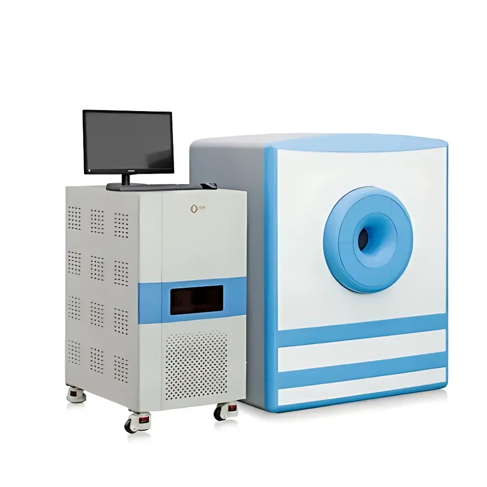
NIUMAG NM42-060H-I Small Animal MRI System for Cell Tracking and In Vivo Imaging
| Brand | NIUMAG |
|---|---|
| Origin | Jiangsu, China |
| Manufacturer Type | Authorized Distributor |
| Country of Origin | China |
| Model | NM42+ |
| Instrument Type | Preclinical MRI System for Rodent Research (Mouse/Rat) |
| Resonance Frequency | ~42 MHz |
| Static Magnetic Field Strength | 1.0 ± 0.05 T |
| Bore Diameter | 60 mm (Dedicated Animal RF Coil) |
| Magnet Type | Permanent Magnet |
| Application Scope | In vivo cell tracking, contrast agent evaluation, tumor modeling, metabolic phenotyping, longitudinal studies in mice and rats |
Overview
The NIUMAG NM42-060H-I is a dedicated benchtop permanent-magnet small animal MRI system engineered for non-invasive, high-contrast in vivo imaging and longitudinal cell tracking in murine models. Operating at a nominal Larmor frequency of 42 MHz—corresponding to a static magnetic field strength of 1.0 ± 0.05 T—the system leverages proton (¹H) signal detection to generate anatomical, functional, and contrast-enhanced images with sub-millimeter spatial resolution. Unlike optical or bioluminescent modalities, this MRI platform provides true three-dimensional soft-tissue contrast without depth limitation or tissue autofluorescence interference. Its design centers on reproducible quantitative imaging of labeled cells—particularly those tagged with superparamagnetic iron oxide nanoparticles (SPIOs)—enabling sensitive detection of cellular migration, engraftment, and retention across organs including brain, liver, spleen, and subcutaneous tumor sites. The system’s permanent magnet architecture ensures stable field homogeneity over time, minimal cryogen dependency, and low operational overhead—making it suitable for core imaging facilities, pharmacology labs, and longitudinal preclinical studies requiring daily or weekly scanning protocols.
Key Features
- Dedicated 60 mm inner-diameter RF coil optimized for mouse-sized subjects (typically 20–30 g), delivering high signal-to-noise ratio (SNR) and uniform excitation across the imaging volume.
- Stable 1.0 T permanent magnet with active shimming capability, achieving field homogeneity better than 10 ppm over a 30 mm DSV (Diameter Spherical Volume).
- Integrated gradient subsystem supporting standard pulse sequences—including spin echo (SE), fast spin echo (FSE), gradient echo (GRE), and inversion recovery (IR)—for T₁-, T₂-, and PD-weighted contrast generation.
- Real-time image reconstruction engine enabling on-console preview and rapid protocol iteration during animal positioning and scan optimization.
- Modular software architecture compatible with DICOM 3.0 export, facilitating integration into institutional PACS environments and multi-modal data correlation workflows (e.g., PET/MRI or histology-MRI registration).
Sample Compatibility & Compliance
The NM42-060H-I supports live, anesthetized mouse and rat imaging under controlled physiological monitoring (optional temperature and respiration gating modules available). It complies with IEC 61000-6-3 (EMC emission standards) and meets mechanical safety requirements per ISO 13485-aligned manufacturing practices. While not FDA-cleared for human use, the system adheres to GLP-relevant documentation standards for preclinical study reporting. All imaging protocols are configurable to align with common regulatory guidance frameworks—including OECD Test Guidelines for repeated-dose toxicity studies and NIH/NCI recommendations for tumor xenograft imaging endpoints. Data integrity is maintained via timestamped acquisition logs and user-accessible audit trails for all sequence modifications and parameter adjustments.
Software & Data Management
The proprietary NIUMAG MRI Console Software (v5.x) provides intuitive workflow navigation, sequence library management, and batch processing tools for ROI-based quantification (e.g., signal intensity decay curves, contrast-to-noise ratio mapping, and voxel-wise T₂* relaxation analysis). Raw k-space data and reconstructed NIfTI/DICOM volumes are stored with embedded metadata—including scanner parameters, animal ID, anesthesia status, and injection timing for contrast agents. The software supports 21 CFR Part 11-compliant user authentication, electronic signatures, and immutable audit logs when deployed in validated GxP environments. Export options include MATLAB-compatible .mat files for advanced statistical modeling and Python-based analysis pipelines using NiBabel or PyDicom libraries.
Applications
- Cell tracking studies: Quantitative localization and persistence assessment of SPIO-labeled immune cells (e.g., macrophages, dendritic cells, CAR-T cells) following intravenous or intratumoral administration.
- Oncology research: Monitoring orthotopic and subcutaneous tumor growth kinetics, necrosis progression, and response to checkpoint inhibitors or anti-angiogenic therapies using dynamic contrast-enhanced (DCE) MRI.
- Neuroinflammation modeling: Detection of microglial activation and blood-brain barrier disruption in EAE, Alzheimer’s, or stroke models via T₂*-weighted susceptibility mapping.
- Metabolic phenotyping: Fat-water separation imaging for visceral adipose tissue quantification in diet-induced obesity or genetic metabolic syndrome models.
- Contrast agent development: In vivo relaxivity (r₁/r₂) characterization and biodistribution profiling of novel nanoparticle formulations under standardized imaging conditions.
FAQ
What is the minimum detectable iron oxide concentration for cell labeling in vivo?
Detection sensitivity depends on labeling efficiency and voxel size; typical thresholds range from 1–5 µg Fe/g tissue under optimized GRE or SWI sequences.
Can the system accommodate longitudinal scans across multiple weeks?
Yes—its passive magnet stability and automated shimming reduce day-to-day variability, supporting robust inter-scan comparison when combined with consistent animal positioning and physiological monitoring.
Is respiratory or cardiac gating supported?
Hardware gating interfaces are available as optional add-ons; software-based retrospective motion correction is included in the base package.
Does the system support diffusion-weighted imaging (DWI)?
Yes—b-values up to 1000 s/mm² are achievable using standard gradient performance specifications.
Are service contracts and application support included?
NIUMAG offers tiered technical support packages, including remote diagnostics, annual performance verification, and on-site application training for new users.


