Phenom Ar-C Argon-Compatible Desktop Scanning Electron Microscope
| Brand | Phenom |
|---|---|
| Origin | Netherlands |
| Model | Phenom Ar-C |
| Instrument Type | Benchtop SEM |
| Electron Source | Cerium Hexaboride (CeB6) |
| Secondary Electron Resolution | 8 nm |
| Maximum Magnification | 200,000× |
| Accelerating Voltage Range | 4.8–20.5 kV |
| Backscattered Electron Resolution | 8 nm |
Overview
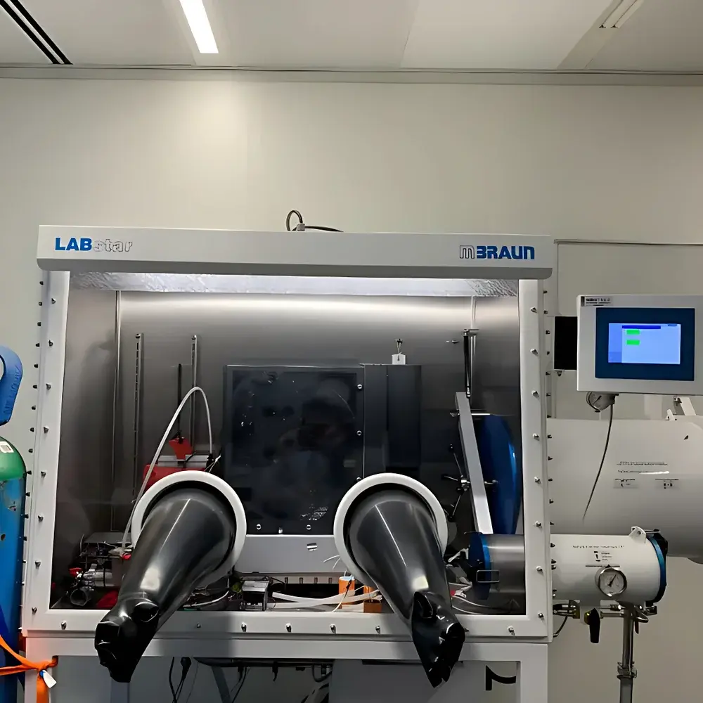
The Phenom Ar-C Argon-Compatible Desktop Scanning Electron Microscope is the world’s first fully functional benchtop SEM engineered for seamless integration and continuous operation inside inert-atmosphere gloveboxes—specifically argon-purged environments with O₂/H₂O levels < 0.5 ppm. Unlike conventional SEMs requiring air-exposed sample transfer, the Phenom Ar-C enables in-situ preparation, imaging, and elemental analysis of air-sensitive materials—including battery cathode/anode powders, alkali metal compounds, organometallic catalysts, and reactive nanomaterials—without exposure to ambient atmosphere. Its design adheres to vacuum-compatible mechanical integrity, low-outgassing material selection, and feedthrough-optimized cabling (Ethernet, USB 2.0, and dedicated power). The system operates on a CeB6 thermionic electron source, delivering stable beam current and high brightness at accelerating voltages from 4.8 kV to 20.5 kV, supporting both morphological contrast (via secondary and backscattered electrons) and quantitative EDS-based compositional analysis—all within the controlled environment of a Class 1000 or better argon glovebox.
Key Features
- Argon-compatible hardware architecture: All external interfaces (power, Ethernet, USB) utilize hermetic feedthroughs rated for ≤0.5 ppm O₂/H₂O environments.
- CeB6 electron source: Offers >1,500-hour typical lifetime, reduced thermal drift, and higher brightness than tungsten filaments—critical for long-duration mapping and automated workflows inside sealed gloveboxes.
- Dual-mode imaging: Standard quad-segment backscattered electron detector (BSD) provides simultaneous topographic and atomic-number contrast; optional secondary electron detector (SED) enhances surface detail at low kV.
- Integrated energy-dispersive X-ray spectroscopy (EDS): Fully embedded silicon drift detector (SDD) with real-time spectrum acquisition, peak identification, and qualitative/quantitative elemental mapping (e.g., Ni-Co-Mn distribution in NMC cathodes).
- Motorized eucentric stage: Enables precise tilt (–15° to +90°), rotation (360° continuous), height (Z), and lateral (x′) motion—essential for 3D reconstruction, cross-sectional imaging, and crystallographic orientation assessment.
- ProSuite software platform: Includes ParticleMetric (ISO 13322-1 compliant particle sizing), PoroMetric (pore size distribution per ASTM D7782), FiberMetric (fiber diameter/alignment analysis), and 3D Roughness Reconstruction modules—all operable without breaking glovebox integrity.
Sample Compatibility & Compliance
The Phenom Ar-C accommodates samples up to 100 mm × 100 mm × 40 mm, with standard scanning area of 50 mm × 50 mm (expandable to 100 mm × 100 mm). It supports conductive and non-conductive specimens—including Li-metal anodes, sodium-ion battery electrodes, MOFs, and pyrophoric metal-organic precursors—without mandatory sputter coating, thanks to low-voltage imaging (down to 4.8 kV) and charge compensation algorithms. System compliance includes adherence to IEC 61000-6-3 (EMC emissions), IEC 61000-6-2 (immunity), and mechanical safety per ISO 12100. For regulated laboratories, ProSuite supports audit trails, user-level access control, and electronic signatures compatible with FDA 21 CFR Part 11 requirements when deployed on validated networked workstations.
Software & Data Management
Acquisition and analysis are unified under ProSuite v5.x, a Windows-based application with Python-programmable interface (PPI). Users can script automated workflows—such as batch particle analysis across multiple fields-of-view, scheduled EDS line scans, or large-area mosaic stitching—using native Python libraries. Image data exports in TIFF (16-bit), JPEG, BMP, and EMSA/ELID formats; spectral data in CSV and standard .eds files. All metadata—including kV, dwell time, working distance, detector mode, and glovebox environmental logs—are embedded in image headers. Data storage options include internal SSD (256 GB), USB 3.0 flash drives, and network-mounted NAS via SMB/CIFS protocols—ensuring traceability and version control aligned with GLP/GMP documentation practices.
Applications
- Battery R&D: In-situ observation of SEI evolution, dendrite growth, and cathode cracking in Li/Na/K-metal cells.
- Heterogeneous catalysis: Morphological and compositional stability assessment of supported Pt, Ru, or Ni nanoparticles under inert handling.
- Quantum materials: Imaging of air-sensitive topological insulators (e.g., Bi₂Se₃) and 2D transition metal dichalcogenides (MoS₂, WSe₂) without oxidation artifacts.
- Pharmaceutical solid-state chemistry: Characterization of moisture-sensitive amorphous dispersions and reactive co-crystals.
- Geochemical reference materials: High-resolution BSE imaging coupled with EDS mapping of trace element zoning in sulfide inclusions under O₂-free conditions.
FAQ
Can the Phenom Ar-C be operated remotely while sealed inside an argon glovebox?
Yes—full remote control via Ethernet is supported, including live imaging, stage navigation, EDS acquisition, and software scripting through the glovebox’s viewport-integrated touchscreen or external workstation.
What vacuum level does the system achieve, and how is it maintained inside the glovebox?
The integrated diaphragm pump achieves base pressure <10⁻¹ Pa in the electron column; no additional roughing pump or turbomolecular pump is required. Vacuum integrity is preserved via welded stainless-steel vacuum path and metal-sealed feedthroughs.
Is EDS quantification accurate under low-kV conditions (e.g., 5 kV)?
Yes—ProSuite applies ZAF matrix corrections and uses k-ratio calibration standards traceable to NIST SRMs; accuracy is validated per ISO 14704 for light-element quantification (C, N, O) in conductive matrices.
How often must the CeB6 filament be replaced in glovebox operation?
Under typical usage (≤4 h/day, 5 kV–10 kV imaging), replacement interval exceeds 1,500 hours; automatic filament sleep mode during idle periods further extends service life.
Does the system support automated large-area imaging (mosaic) across multiple tiles?
Yes—MosaicScan module enables programmable grid acquisition up to 100 × 100 tiles (7680 × 4800 px per tile), with sub-pixel image registration and seam-free stitching.


