TESCAN VEGA COMPACT Tungsten-Filament Scanning Electron Microscope
| Brand | TESCAN |
|---|---|
| Origin | Czech Republic |
| Manufacturer | TESCAN s.r.o. |
| Type | Imported Instrument |
| Model | TESCAN VEGA COMPACT |
| Electron Source | Tungsten Filament |
| Secondary Electron Resolution | 3.0 nm @ 30 kV |
| Magnification Range | 2× to 1,000,000× |
| Accelerating Voltage | 200 V – 30 kV (continuously adjustable) |
| Backscattered Electron Resolution | 3.5 nm @ 30 kV |
Overview
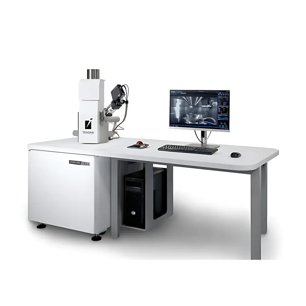
The TESCAN VEGA COMPACT is a high-performance, entry-level tungsten-filament scanning electron microscope (SEM) engineered for routine laboratory applications in materials science, quality assurance, failure analysis, and academic research. Designed on the proven platform of the VEGA series, it integrates robust electron optics with a compact, space-efficient architecture—ideal for laboratories requiring reliable SEM capabilities without the complexity or cost of field-emission systems. Its operation relies on thermionic emission from a tungsten hairpin filament, delivering stable beam current and consistent imaging performance across a wide accelerating voltage range (200 V–30 kV). The instrument employs a fully electromagnetic lens column with optimized probe-forming optics, enabling high signal-to-noise imaging at low kV for surface-sensitive contrast and enhanced topographic fidelity. Unlike conventional SEMs relying on mechanical apertures, the VEGA COMPACT utilizes TESCAN’s proprietary aperture-free optical path and In-flight Beam Tracing™ technology, ensuring dynamic beam alignment and automatic optimization of focus, stigmation, and scan parameters—critical for reproducible results in multi-user environments.
Key Features
- Fully integrated TESCAN Essence™ software platform unifying real-time SEM imaging and energy-dispersive X-ray spectroscopy (EDS) analysis within a single interface—eliminating workflow fragmentation between morphological and compositional characterization.
- Aperture-free optical design with In-flight Beam Tracing™ for automated, real-time electron beam diagnostics and condition optimization—reducing setup time and operator dependency.
- Wide Field Optics™ enabling true 2× minimum magnification, allowing full-sample navigation without auxiliary optical cameras or stage repositioning.
- 5-axis motorized eucentric stage (X/Y: ±40 mm; Z: 50 mm; tilt: −80° to +80°; rotation: 360° continuous) with precise center-of-rotation alignment for accurate tilt-series acquisition and cross-sectional imaging.
- Essence™ 3D collision avoidance model simulating detector and pole-piece positions during stage motion—preventing hardware damage during automated workflows or novice operation.
- Optional vacuum buffer unit significantly reduces mechanical pump runtime, lowering power consumption and extending pump service intervals—supporting sustainable lab operations.
Sample Compatibility & Compliance
The VEGA COMPACT accommodates samples up to 145 × 145 mm in planar dimension and 81 mm in height, with a maximum weight of 1 kg. Its versatile chamber configuration supports conductive, semi-conductive, and non-conductive specimens—with optional low-vacuum mode or carbon/gold sputter coating for charge mitigation. The system complies with IEC 61000-6-3 (EMC emissions) and IEC 61000-6-2 (immunity), and meets CE marking requirements for laboratory equipment. Software features—including audit trail logging, user access levels, parameter locking, and timestamped session records—support GLP and ISO/IEC 17025-aligned documentation practices. Optional CORAL™ module enables correlative light-electron microscopy (CLEM) workflows compliant with life science imaging standards.
Software & Data Management
Powered by TESCAN Essence™—a modular, role-based SEM control suite—the VEGA COMPACT delivers intuitive yet powerful data acquisition and processing. Core capabilities include: measurement and tolerance analysis tools; histogram/LUT-based contrast enhancement; SharkSEM™ remote control (basic); automatic stitching (mosaic imaging); offline data processing via TESCAN Flow™; and customizable UI layouts tailored to user expertise or application focus (e.g., metallurgy, geology, polymers). All acquired images and spectra are stored in vendor-neutral TIFF/EMD formats, with metadata embedded per ASTM E1558 and ISO 16700 standards. Session logs support traceability for QA/QC audits, while optional FDA 21 CFR Part 11-compliant modules provide electronic signature, change control, and secure archive functions.
Applications
The VEGA COMPACT serves as a primary analytical tool in diverse domains: microstructural evaluation of metals, ceramics, and composites; particle size and morphology analysis in pharmaceuticals and catalysts; fracture surface examination in failure analysis; coating thickness and uniformity assessment; semiconductor package inspection; and educational training in electron microscopy fundamentals. Its low-kV imaging capability (<5 kV) enables high-resolution surface detail on beam-sensitive polymers and biological tissues (with appropriate preparation), while its EDS integration supports quantitative elemental mapping and phase identification per ASTM E1508 and ISO 22309 protocols.
FAQ
What is the typical working distance range for optimal resolution?
The standard working distance is 10–25 mm, with resolution specifications (3.0 nm SE, 3.5 nm BSE) measured at 10 mm WD and 30 kV.
Is EDS hardware included as standard or optional?
A fully integrated EDS detector is available as a factory-configured option; system compatibility requires the Essence™ EDS module license and dedicated detector port.
Can the VEGA COMPACT operate in low vacuum mode?
Yes—optional variable pressure (VP) mode supports partial pressures up to 130 Pa, enabling imaging of non-conductive or hydrated samples without metal coating.
What vacuum level does the system achieve in high vacuum mode?
The base pressure is ≤1 × 10⁻³ Pa, maintained by a turbomolecular pump backed by an oil-free diaphragm pump.
Does the software support batch image acquisition and automated report generation?
Yes—via TESCAN Flow™ and custom macro scripting, users can define multi-location acquisition routines and export annotated reports in PDF or HTML format with embedded metadata.


