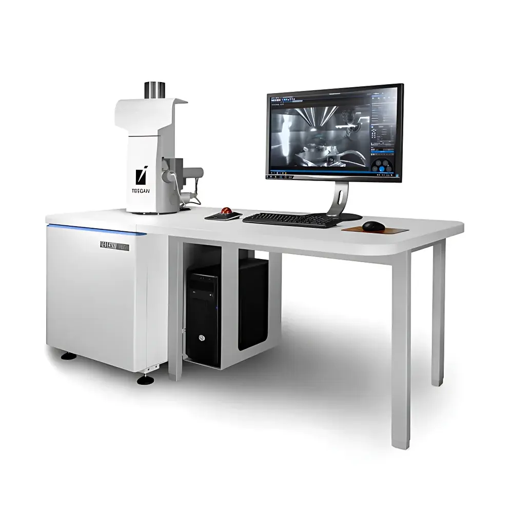
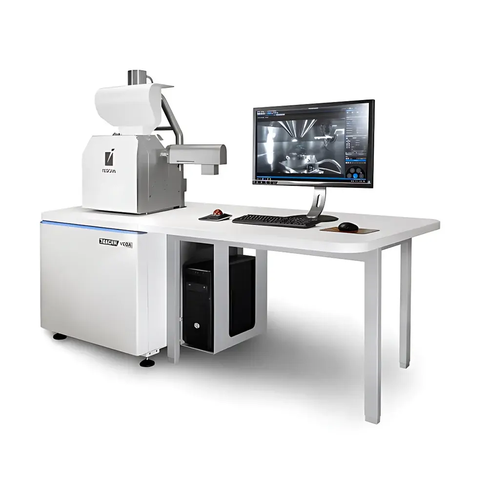
TESCAN VEGA Tungsten-Filament Scanning Electron Microscope
| Brand | TESCAN |
|---|---|
| Origin | Czech Republic |
| Manufacturer | TESCAN ORSAY HOLDING a.s. |
| Type | VEGA |
| Electron Source | Tungsten (W) Filament |
| Secondary Electron Resolution | 3.0 nm @ 30 kV |
| Magnification Range | 2× to 1,000,000× |
| Accelerating Voltage | 0.2–30 kV (continuously adjustable) |
| Backscattered Electron Resolution | 3.5 nm @ 30 kV |
Overview
The TESCAN VEGA is the fourth-generation tungsten-filament scanning electron microscope (SEM), engineered for high reliability, operational efficiency, and seamless integration of imaging and microanalysis in routine and research-grade laboratories. Based on a field-proven thermionic electron source, the VEGA delivers consistent beam current stability and long filament lifetime—critical for high-throughput quality control, failure analysis, and materials characterization where cost-of-ownership and uptime are key performance indicators. Its electron optical column features a fully electromagnetic lens system with real-time In-Flight Beam Tracing™ technology, eliminating the need for mechanical aperture alignment or manual gun tuning during operation. This architecture ensures rapid, reproducible transition between imaging and analytical modes without compromising resolution or signal fidelity. The system operates under standard high-vacuum conditions but includes SingleVac™ mode as standard—a differential pumping configuration enabling direct observation of non-conductive, hydrated, or beam-sensitive specimens without sputter coating or cryo-preparation.
Key Features
- Essence™ Integrated Software Platform: A unified graphical interface combining SEM imaging, EDS elemental mapping, and quantitative analysis within a single scan window—no context switching or external software modules required.
- Wide Field Optics™: Enables true low-magnification navigation down to 2× with distortion-free, high-depth-of-field imaging directly in the SEM viewport—replacing conventional optical navigation cameras and streamlining region-of-interest targeting.
- Mechanical Aperture-Free Design: Eliminates physical apertures from the beam path, reducing contamination risk and maintenance frequency while improving signal-to-noise ratio across all operating voltages.
- 3D Anti-Collision Model: Real-time visualization of detector positions, stage coordinates, and sample geometry within the chamber—preventing hardware collisions during automated tilt, rotation, or multi-detector workflows.
- SingleVac™ Mode: Standard vacuum configuration supporting variable-pressure imaging of insulating or delicate samples (e.g., polymers, biological tissues, ceramics) at pressures up to 100 Pa, without conductive coating.
- Modular Detector Architecture: Supports simultaneous installation of Everhart-Thornley SE, retractable scintillator BSE, cathodoluminescence (CL), cooled BSE, and optional Raman spectroscopy integration via dedicated ports and synchronized acquisition triggers.
Sample Compatibility & Compliance
The VEGA accommodates a broad range of specimen types—from bulk metallic alloys and geological sections to fragile biological specimens, thin films, and nanocomposites. Its SingleVac™ capability complies with ASTM E1508 and ISO 16700 guidelines for non-conductive sample evaluation, while its stable beam performance meets GLP/GMP documentation requirements for regulated environments. All EDS-based quantification workflows adhere to IUPAC recommendations for standardless analysis and support traceable calibration using certified reference materials (CRMs). The system’s embedded audit trail, user-access logging, and parameter versioning align with FDA 21 CFR Part 11 readiness when configured with appropriate IT infrastructure and procedural controls.
Software & Data Management
Essence™ employs a modular, Python-scriptable architecture that exposes full instrument control—including stage motion, beam parameters, detector gating, and image acquisition—to custom automation. Predefined workflows (e.g., particle sizing, line scans, area mapping) can be saved, shared, and re-executed with parameter inheritance. Offline data processing is supported via TESCAN Flow™, enabling batch image enhancement, spectral deconvolution, phase identification, and 3D surface reconstruction without occupying the microscope. Measurement tools include calibrated distance/angle/area annotation, histogram-based thresholding, LUT adjustment, and statistical reporting exportable to CSV, TIFF, or HDF5 formats. Optional modules include SharkSEM™ for remote diagnostics, CORAL™ for life science-specific contrast optimization, and automated mosaic stitching for large-area survey imaging.
Applications
The VEGA serves as a foundational platform across multiple domains: metallurgical QC labs use it for inclusion analysis and fracture surface evaluation per ASTM E1245; semiconductor fabs apply it for defect review and cross-section metrology; academic researchers employ it for catalyst morphology studies, battery electrode porosity quantification, and geoscience texture analysis; and polymer engineers rely on its low-kV imaging for surface topography and filler dispersion assessment. Its compatibility with EBSD pre-tilt stages and synchronized EDS-EBSD acquisition enables crystallographic phase mapping in multiphase alloys and mineral assemblages—supporting ISO/IEC 17025-compliant testing protocols.
FAQ
What vacuum modes does the VEGA support?
The system operates in high vacuum (HV) mode for maximum resolution and in SingleVac™ mode for variable-pressure imaging up to 100 Pa—both selectable via software without hardware modification.
Can the VEGA perform automated particle analysis?
Yes—using the optional Particle Analysis module, users define morphological criteria (size, aspect ratio, circularity) and execute batch measurements across multiple fields, generating statistically robust reports compliant with ISO 9276 standards.
Is EDS integration hardware-agnostic?
No—the VEGA ships with native support for TESCAN’s integrated Essence™ EDS platform; third-party detectors require custom driver development and are not officially supported for quantitative workflows.
How is beam alignment handled?
Automatic filament heating, gun alignment, and stigmator correction are executed via one-click routines; In-Flight Beam Tracing™ continuously monitors beam trajectory and adjusts lens excitations in real time.
Does the system support remote operation and diagnostics?
Yes—SharkSEM™ enables secure LAN/WAN-based remote control, live session sharing, and predictive diagnostics via encrypted TLS communication, compatible with enterprise IT security policies.


