Thermo Fisher Scientific Invitrogen E-Gel Imager Gel Documentation System
| Brand | Thermo Fisher Scientific |
|---|---|
| Origin | USA |
| Manufacturer Type | Original Equipment Manufacturer (OEM) |
| Import Status | Imported |
| Model | E-Gel |
| Instrument Category | Standard Gel Imaging System |
| CCD Resolution | 1,296 (H) × 964 (V) |
| Bit Depth | 12-bit |
| Dynamic Range | 56.77 dB |
| CCD Sensor Size | 11 × 14 cm |
| Lens | F/1.4 Optimized Fixed-Focus Lens with 6–12 mm Optical Focal Length |
Overview
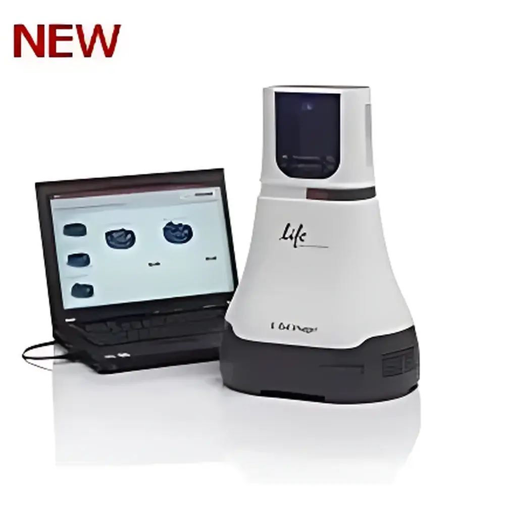
The Thermo Fisher Scientific Invitrogen E-Gel Imager is a compact, benchtop gel documentation system engineered for reliable, high-fidelity imaging of DNA and RNA electrophoresis gels—primarily E-Gel® pre-cast agarose gels, but also compatible with standard hand-poured agarose and polyacrylamide gels. It employs a scientific-grade monochrome CCD sensor optimized for UV transillumination (254 nm and 302 nm) and blue-light excitation (for SYBR® Safe, GelGreen®, and other non-UV nucleic acid stains), delivering consistent signal capture without thermal noise accumulation. Unlike large shared-platform systems requiring dedicated darkroom space and multi-user scheduling, the E-Gel Imager integrates optics, illumination control, and image acquisition into a single self-contained unit—designed specifically for individual lab users who require rapid, reproducible gel documentation without compromising analytical integrity.
Key Features
- 1.3-megapixel monochrome CCD sensor with 12-bit analog-to-digital conversion ensures high-resolution digitization and quantifiable grayscale linearity across the full dynamic range (56.77 dB).
- F/1.4 high-aperture fixed-focus lens (6–12 mm focal length) provides uniform field illumination and sharp edge-to-edge resolution across the 11 × 14 cm active imaging area—optimized for standard E-Gel cassettes and mini-gel formats.
- Dedicated UV and white-light transillumination modes with adjustable intensity control enable safe, low-photobleaching imaging of ethidium bromide, SYBR® dyes, and protein stains (e.g., Coomassie Blue, Silver Stain).
- Integrated USB 2.0 interface enables direct connection to Windows-based workstations; no external frame grabber or driver installation required beyond standard Thermo Fisher software suite.
- Compact footprint (W × D × H ≈ 32 × 35 × 30 cm) and lightweight design (<8 kg) allow flexible placement on standard laboratory benches, fume hoods, or mobile carts—ideal for core facilities with space constraints or teaching labs requiring portability.
Sample Compatibility & Compliance
The E-Gel Imager supports all E-Gel® pre-cast agarose electrophoresis cassettes (including E-Gel® EX, E-Gel® iBase-compatible, and E-Gel® CloneWell formats), as well as conventional 7 × 10 cm and 10 × 10 cm hand-cast agarose and native/polyacrylamide gels. It complies with IEC 61010-1:2010 safety standards for electrical equipment used in laboratory environments. While not certified for GLP/GMP production environments, its hardware architecture and software logging capabilities support audit-ready workflows under ISO/IEC 17025-accredited testing laboratories when paired with documented SOPs and user-access controls. UV exposure time is programmable and logged per acquisition—supporting traceability requirements aligned with internal quality assurance protocols.
Software & Data Management
The system operates exclusively with Thermo Fisher’s GelQuant Express™ software (v3.x or later), a validated, standalone application for image acquisition, background subtraction, band detection, molecular weight estimation, and relative quantification. All image metadata—including exposure time, lamp mode, lens position, and user ID—are embedded in TIFF and PNG exports. Software supports batch processing of multiple gels, export of calibrated intensity values (arbitrary units), and generation of publication-ready figures compliant with journal submission guidelines (e.g., Nature, PNAS). Audit trail functionality records user actions, timestamps, and parameter modifications—facilitating compliance with FDA 21 CFR Part 11 when deployed in regulated research settings with appropriate administrative controls.
Applications
- Routine verification of PCR amplification products, restriction digests, and cloning constructs in molecular biology workflows.
- Quantitative comparison of gene expression levels via RT-PCR or Northern blotting using densitometric normalization against loading controls.
- Assessment of DNA fragmentation patterns in apoptosis assays and genotoxicity screening.
- Validation of CRISPR-Cas9 editing efficiency by T7E1 or Surveyor nuclease assays.
- Teaching laboratory instruction in undergraduate genetics and biochemistry courses—where rapid turnaround, intuitive interface, and minimal training overhead are critical.
FAQ
Is the E-Gel Imager compatible with non-E-Gel® agarose gels?
Yes—it accepts standard mini-gel formats up to 10 × 10 cm, provided they fit within the 11 × 14 cm imaging field and are placed on the built-in UV-transparent tray.
Does it support fluorescence imaging with SYBR® Gold or GelRed®?
Yes, when used with optional blue-light LED transilluminator accessories (sold separately); native UV modes are optimized for EtBr and SYBR® Safe.
Can images be exported in formats suitable for journal submission?
Yes—TIFF (uncompressed, 12-bit), PNG, and JPEG outputs are supported; GelQuant Express allows annotation-free export at user-defined DPI (up to 600 dpi) and color space (grayscale or pseudocolor LUTs).
Is remote operation or network deployment possible?
No—the system connects via USB 2.0 only and does not support Ethernet, Wi-Fi, or multi-user server deployment; it is designed for single-user, local workstation use.
What maintenance is required for long-term optical performance?
Annual calibration of UV lamp intensity and CCD dark-current offset is recommended; Thermo Fisher offers certified service plans including sensor diagnostics and lens cleaning validation.


