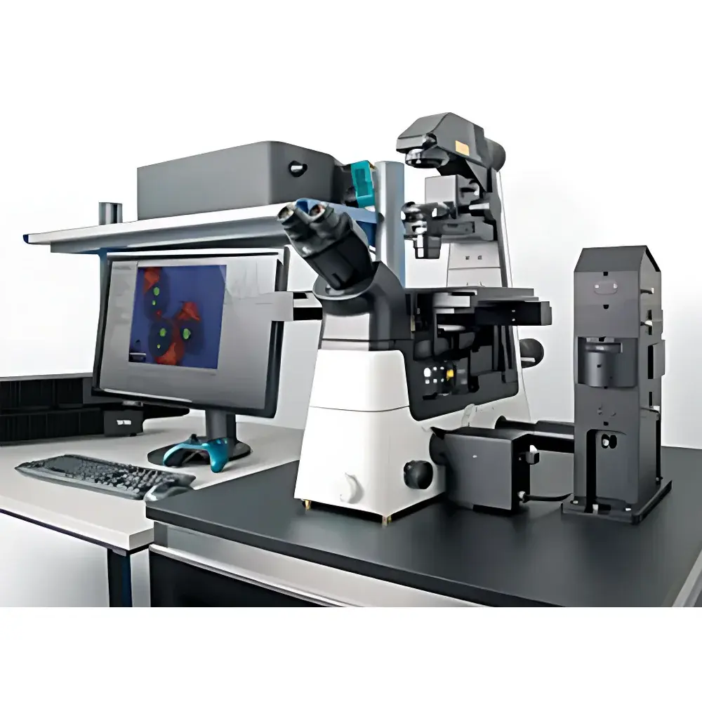
WITec Alpha 300RI Inverted Confocal Raman Imaging System
| Brand | WITec |
|---|---|
| Origin | Germany |
| Model | Alpha 300RI |
| Instrument Type | Confocal Micro-Raman Spectrometer |
| Spectral Range | 90–9000 cm⁻¹ |
| Spectral Resolution | ≤0.2 cm⁻¹ |
| Spatial Resolution | Lateral 350 nm, Axial 800 nm |
| Low-Wavenumber Cutoff | 10 cm⁻¹ |
| Spectral Reproducibility | ≤±0.02 cm⁻¹ |
Overview
The WITec Alpha 300RI Inverted Confocal Raman Imaging System is an engineered platform for high-fidelity, non-invasive chemical mapping of heterogeneous samples in three dimensions. Unlike conventional upright confocal Raman microscopes, the Alpha 300RI integrates a fully inverted optical architecture—co-aligning a research-grade inverted microscope with a high-throughput confocal Raman spectrometer via fiber-coupled optics. This design preserves the core principles of confocal detection (axial sectioning via pinhole rejection, diffraction-limited lateral resolution) while enabling unobstructed access to the sample plane from below. The system operates on spontaneous Raman scattering, where monochromatic laser excitation induces inelastic photon scattering; spectral shifts (cm⁻¹) are measured with sub-0.02 cm⁻¹ reproducibility across the full 90–9000 cm⁻¹ range. Its inverted configuration is particularly suited for live-cell imaging in aqueous environments, large-area substrates (e.g., wafers, tissue sections), and dynamic in situ experiments requiring environmental chambers—without compromising optical stability or alignment fidelity.
Key Features
- Inverted optical layout enables direct integration with standard inverted microscope accessories—including incubation chambers, micromanipulators, perfusion systems, and motorized stage modules—without optical path interference.
- Confocal Raman imaging with lateral resolution down to 350 nm and axial resolution of 800 nm, achieved through precision pinhole alignment and aberration-corrected UHTS spectrograph optics.
- Fiber-coupled Ultra-High-Throughput Spectrograph (UHTS) delivering >70% optical throughput across UV–VIS–NIR-optimized configurations, optimized for low-signal applications such as single-cell biochemistry or carbon nanomaterial characterization.
- Motorized XY stage (110 × 70 mm travel) compatible with standard Petri dishes, multi-well plates, and custom sample holders; supports automated multi-region acquisition and stitching of large-area hyperspectral datasets.
- Full compatibility with complementary contrast modalities: brightfield, phase contrast, differential interference contrast (DIC), and optional epi-fluorescence—enabling correlative Raman–fluorescence imaging (e.g., DAPI-stained nuclei overlaid with nucleic acid Raman signatures).
- Non-destructive, label-free chemical imaging: no staining, fixation, or metal coating required; preserves native sample integrity for longitudinal studies under physiological conditions.
Sample Compatibility & Compliance
The Alpha 300RI accommodates diverse sample formats—including liquid-phase biological specimens (e.g., cultured cells in growth medium), thick tissue slices (>100 µm), geological thin sections, polymer composites, and semiconductor wafers—without reconfiguration. Its inverted geometry eliminates top-access constraints inherent to upright systems, permitting simultaneous optical access from above (for fluorescence excitation or manipulation) and below (for transmitted illumination or stage-mounted peripherals). The system meets essential regulatory expectations for analytical instrumentation used in GLP-compliant laboratories: software supports audit-trail logging, user-access controls, and electronic signature functionality aligned with FDA 21 CFR Part 11 requirements. All spectral acquisitions adhere to ISO/IEC 17025 traceability frameworks when calibrated using NIST-traceable standards (e.g., silicon, cyclohexane).
Software & Data Management
Control, acquisition, and analysis are unified within WITec’s proprietary Project software suite—designed specifically for multidimensional Raman data handling. The platform supports real-time spectral preview during acquisition, automated background subtraction (polynomial or morphological), multivariate analysis (PCA, cluster analysis), and quantitative mapping of molecular species via peak intensity ratioing or curve-fitting (e.g., Lorentzian/Gaussian deconvolution). Hyperspectral cubes (x, y, λ) are stored in vendor-neutral HDF5 format, ensuring interoperability with third-party tools (MATLAB, Python SciPy, HyperTools). Data provenance—including laser power, integration time, grating selection, and objective magnification—is embedded in metadata headers. For regulated environments, the software provides configurable report templates compliant with ASTM E1847 (standard practice for Raman spectral data exchange) and USP (Raman spectroscopy in pharmaceutical analysis).
Applications
- Life Sciences: Subcellular mapping of lipid droplets, protein conformational states, and nucleic acid distribution in live adherent cells—correlated with fluorescence markers without photobleaching artifacts.
- Materials Science: Strain mapping in 2D materials (graphene, MoS₂), phase identification in battery electrode cross-sections, and degradation analysis of polymer blends under thermal cycling.
- Geosciences: In situ mineral identification and fluid inclusion analysis in polished thin sections, leveraging low-wavenumber sensitivity (<100 cm⁻¹) for lattice mode detection.
- Pharmaceutical Development: Polymorph screening in tablet coatings, API-excipient interaction studies, and counterfeit drug authentication via spectral fingerprint matching against reference libraries.
- Nanotechnology: Plasmon-enhanced Raman mapping of gold nanoparticle assemblies, spatially resolved stress/strain fields in nanocomposites, and defect quantification in CVD-grown films.
FAQ
Can the Alpha 300RI perform true 3D Raman tomography?
Yes—the system acquires optically sectioned spectra at defined Z-steps using piezo-driven objective focus or motorized stage control, reconstructing volumetric chemical maps with axial resolution of 800 nm.
Is fluorescence background suppression supported?
Yes—hardware-based notch and edge filters are integrated per laser line; software also implements adaptive background modeling and baseline correction algorithms optimized for fluorescent biological matrices.
What laser options are available?
Standard configurations include 532 nm, 633 nm, and 785 nm diode-pumped solid-state lasers; UV (355 nm) and NIR (1064 nm) options are available for resonance enhancement or reduced photodamage.
Does the system support automated multi-point analysis across multiple samples?
Yes—via programmable stage coordinates, grid-based region-of-interest (ROI) definition, and batch script execution, enabling unattended acquisition across dozens of wells or slides.
Can existing inverted microscope components be reused?
Yes—the Alpha 300RI is designed as a modular add-on; standard Nikon Eclipse Ti or Zeiss Axio Observer bodies integrate directly without modification to the base microscope’s optical train or mechanical interface.
Related Products


")