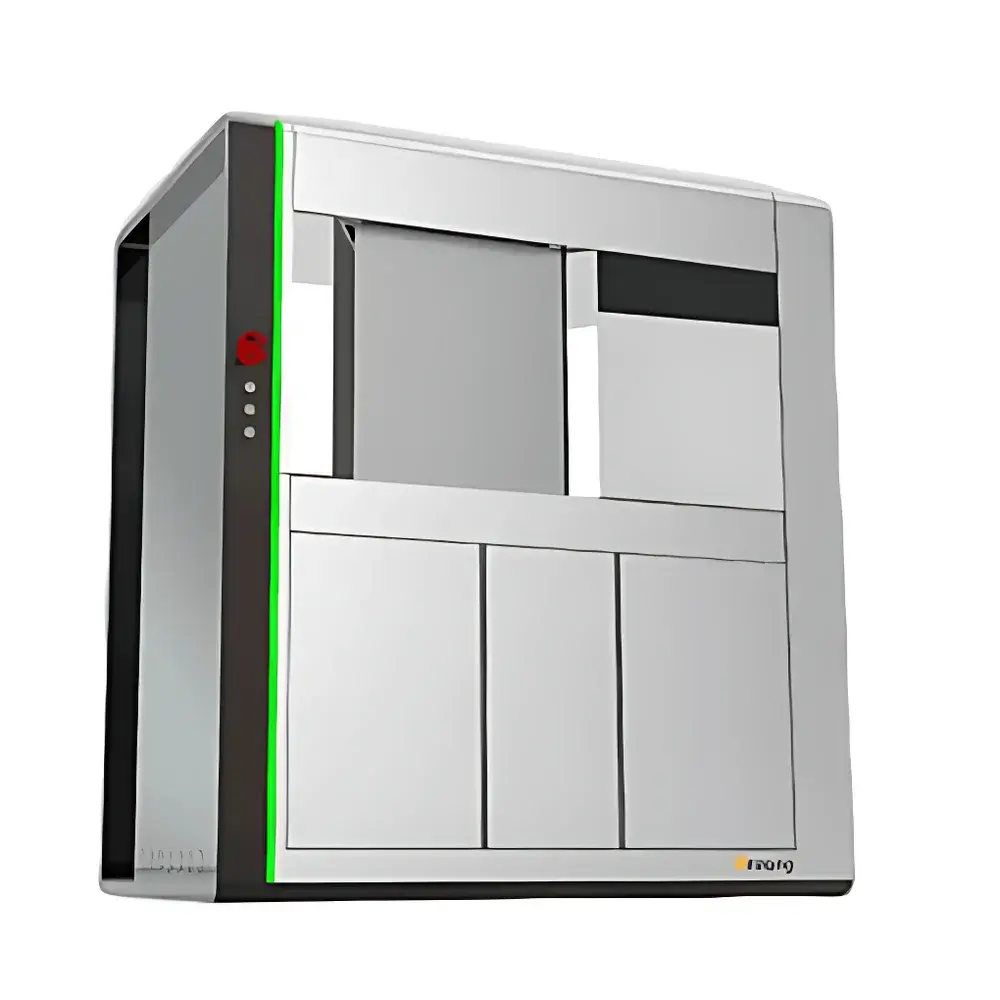
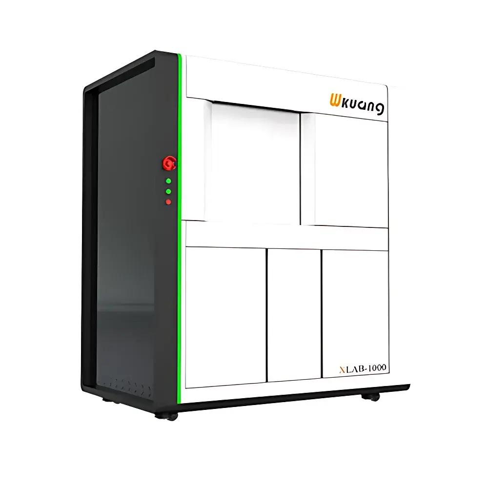
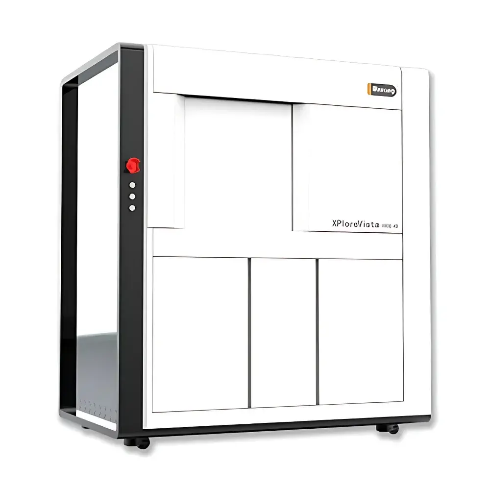
Wkuang XPloreVista 1000 4D Industrial Micro-CT System
| Brand | Wkuang |
|---|---|
| Origin | Jiangsu, China |
| Manufacturer Type | Authorized Distributor |
| Origin Category | Domestic (China) |
| Model | XPloreVista 1000 4D |
| Pricing | Upon Request |
| X-ray Source | Maintenance-Free Integrated Microfocus Tube |
| Detector Pixel Size | 6.5 µm |
| Imaging Modes | Absorption Contrast, Phase Contrast (Propagation-Based), Dynamic 4D Acquisition |
| Software Platform | X-VISON v3.2 (Reconstruction, Artifact Correction, In Situ Data Synchronization) |
| Compliance | ASTM E1441, ISO 15747, IEC 62495 |
Overview
The Wkuang XPloreVista 1000 4D is a high-precision industrial micro-computed tomography (micro-CT) system engineered for non-destructive, volumetric characterization of materials at sub-micron spatial resolution. Operating on the principle of cone-beam X-ray computed tomography, it reconstructs 3D internal structures from hundreds of 2D projection images acquired during sample rotation. Unlike conventional absorption-only CT systems, the XPloreVista 1000 4D integrates propagation-based phase contrast imaging (PCI), enabling enhanced edge delineation and improved soft-tissue or low-Z material contrast—particularly critical for composites, ceramics, polymers, and biological scaffolds where elemental absorption coefficients are nearly identical. Its design targets research-intensive environments including university laboratories, national institutes, and R&D centers conducting advanced failure analysis, porosity quantification, fiber orientation mapping, and time-resolved structural evolution studies.
Key Features
- Maintenance-free integrated microfocus X-ray source (XLAB-1000 platform): No consumables, no filament replacement, stable output over >10,000 hours; optimized for long-duration in situ experiments.
- Ultra-high spatial resolution: 6.5 µm detector pixel pitch enables effective voxel sizes down to 0.8 µm (with geometric magnification), supporting quantitative analysis of fine microstructural features such as grain boundaries, microcracks, and nanopore networks.
- Dual-contrast imaging capability: Simultaneous acquisition and post-processing support for both absorption contrast and propagation-based phase contrast, significantly improving signal-to-noise ratio in low-contrast samples without requiring synchrotron radiation.
- Modular in situ chamber architecture: Standardized flange interfaces and ≥120 mm internal cavity diameter allow seamless integration with third-party or Wkuang-developed mechanical, thermal, and electrochemical stages—including tensile/compression rigs (±5 kN), high-temperature furnaces (up to 1200 °C), and corrosion cells.
- Real-time 4D acquisition engine: Hardware-synchronized trigger logic ensures precise temporal alignment between motion control signals and X-ray exposure, enabling reliable reconstruction of dynamic processes across thousands of time points.
Sample Compatibility & Compliance
The XPloreVista 1000 4D accommodates samples up to Ø150 mm × H200 mm (standard configuration), with optional extended-stage variants supporting larger geometries. It complies with international standards governing industrial CT methodology and data integrity, including ASTM E1441 (Standard Practice for Computed Tomography), ISO 15747 (Non-destructive testing — Industrial computed tomography — General principles), and IEC 62495 (Safety requirements for X-ray equipment). All system firmware and X-VISON software support audit trail logging, user access control, and electronic signature capabilities aligned with GLP/GMP documentation workflows. While not FDA-cleared for clinical use, its reconstruction pipeline adheres to ISO/IEC 17025 traceability principles for metrological validation of dimensional measurements.
Software & Data Management
X-VISON v3.2 is a native cross-platform application (Windows/Linux) delivering end-to-end workflow automation—from scan planning and real-time preview to GPU-accelerated FDK and iterative reconstruction (SART, OS-SART). Key modules include:
- Adaptive scanning scheduler: Supports limited-angle, region-of-interest (ROI), and time-resolved 4D protocols with automatic dose optimization.
- Advanced artifact mitigation suite: Includes ring artifact suppression via frequency-domain filtering, beam hardening correction using dual-energy modeling, and motion blur compensation through projection-domain registration.
- Quantitative analysis toolkit: Provides ISO 12799-compliant pore network extraction, grain size distribution (ASTM E112), wall thickness mapping, and defect classification based on EN 13445 Annex G criteria.
- API-enabled interoperability: RESTful interface supports integration with LIMS, MES, and Python-based analytical pipelines (e.g., scikit-image, PyTorch for AI-assisted segmentation).
Applications
This system serves mission-critical roles across multiple domains:
- Aerospace: Inspection of additively manufactured turbine blades for lack-of-fusion defects, residual stress-induced cracking, and powder remnant quantification.
- Energy materials: In operando tracking of lithium dendrite growth in solid-state batteries, sintering densification kinetics in SOFC anodes, and thermal fatigue damage accumulation in nuclear fuel cladding.
- Geosciences: Pore-throat network modeling in shale cores under confining pressure, CO₂ saturation dynamics in sandstone reservoir analogs.
- Biomaterials: 4D monitoring of scaffold degradation in simulated physiological environments, osteointegration progression in porous titanium implants.
- Electronics: Void detection in solder joints, wire bond integrity assessment, and delamination mapping beneath encapsulants.
FAQ
Does the XPloreVista 1000 4D require external cooling or HVAC infrastructure?
No—it employs air-cooled X-ray source and detector modules; ambient temperature stability within 20–25 °C is recommended for optimal detector dark current performance.
Can X-VISON export reconstructed volumes in standard DICOM or HDF5 formats?
Yes—native export to DICOM (for PACS compatibility), NIfTI, TIFF stacks, and HDF5 with metadata embedding (including acquisition parameters, calibration logs, and user annotations).
Is phase contrast imaging available without synchrotron radiation?
Yes—propagation-based phase contrast is enabled by source–sample–detector distance tuning and does not require coherence-enhancing optics or synchrotron beamlines.
What is the maximum frame rate achievable in dynamic 4D mode?
Up to 30 projections per second (at 2048×2048 resolution, binning 2×2), scalable via hardware ROI selection and GPU-accelerated on-the-fly reconstruction.
Does the system support automated batch processing for multi-sample studies?
Yes—X-VISON includes scriptable batch reconstruction and analysis queues with error handling, email notification, and QC flagging for outlier datasets.