Aperio GT450 Digital Pathology Slide Scanner
| Brand | Leica |
|---|---|
| Origin | Germany |
| Manufacturer Type | Authorized Distributor |
| Import Status | Imported |
| Model | Aperio GT450 |
| Electron Source | Cerium Hexaboride (CeB₆) |
| Scan Speed | 32 s per 15 mm × 15 mm area at 40× |
| Throughput | 81 slides/hour |
| Color Calibration | ICC-compliant medical-grade color profile (DICOM GSDF & sRGB aligned) |
| Optical System | Custom Leica 40× apochromatic objective with real-time autofocus |
| Intended Use | For Research Use Only (RUO) |
Overview
The Aperio GT450 Digital Pathology Slide Scanner is a high-throughput, clinical-grade whole slide imaging (WSI) platform engineered by Leica Biosystems for research laboratories requiring robust, reproducible digitization of histopathological specimens. Unlike transmission electron or scanning electron microscopes—which rely on electron beam interaction with vacuum-mounted conductive samples—the GT450 operates on brightfield optical microscopy principles: it employs high-resolution digital line-scan cameras coupled with precision motorized stages and Leica’s proprietary apochromatic 40× objective to capture gigapixel-scale images from standard glass-mounted H&E, IHC, and special stain slides. Designed explicitly for RUO applications, the system delivers diagnostic-relevant spatial fidelity, chromatic accuracy, and z-stack consistency without requiring vacuum chambers, metal coating, or electron optics. Its architecture prioritizes workflow integration in centralized pathology cores, enabling unattended scanning across heterogeneous slide batches while maintaining traceable focus stability and illumination uniformity per ISO 15189–aligned operational protocols.
Key Features
- High-speed continuous loading: Integrated rack-based feeder accommodates up to 450 standard 1″ × 3″ glass slides; automatic slide recognition and barcode validation ensure error-free batch processing.
- Real-time dynamic autofocus: Proprietary Leica optical feedback loop continuously adjusts Z-position during scanning—compensating for cover-slip thickness variation (0.13–0.17 mm), tissue topography, and thermal drift—without reliance on pre-scan focus maps.
- Medical-grade color fidelity: Factory-calibrated LED illumination paired with ICC v4 profiles compliant with DICOM Grayscale Standard Display Function (GSDF) ensures cross-monitor consistency across PACS, review workstations, and telepathology platforms.
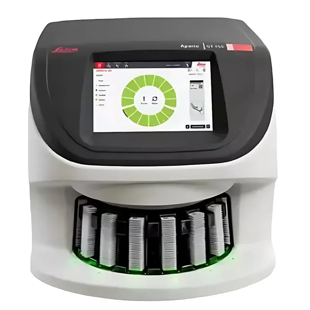
- Touch-integrated control interface: Embedded 12.1″ capacitive touchscreen eliminates dependency on external PCs; supports direct annotation, region-of-interest tagging, and immediate thumbnail preview without software installation.
- Robust mechanical architecture: Precision-ground granite stage base, vibration-damped optical path, and sealed optical chamber minimize particulate ingress—meeting ISO 14644-1 Class 8 cleanroom compatibility requirements for lab environments.
Sample Compatibility & Compliance
The Aperio GT450 accepts conventional formalin-fixed paraffin-embedded (FFPE) and frozen-section slides mounted on 1-mm-thick microscope glass, with or without coverslips (0.13–0.17 mm). It supports standard staining modalities including hematoxylin and eosin (H&E), immunohistochemistry (IHC), fluorescence multiplex (with optional filter wheel), and histochemical stains (e.g., PAS, Masson’s trichrome). While labeled “For Research Use Only,” the system conforms to IEC 61000-6-3 (EMC emissions), IEC 61000-6-2 (immunity), and UL 61010-1 safety standards. Its audit trail functionality—including timestamped operator logins, scan parameter logs, and image hash generation—supports GLP-aligned documentation practices. Though not FDA-cleared for primary diagnosis, its image quality validation framework aligns with CAP checklist ANP.42100 and ISO/IEC 17025 clause 7.7 on measurement uncertainty assessment.
Software & Data Management
The scanner ships with Aperio ImageScope v13.3 and eSlide Manager v14.2, both supporting DICOM WSI Part 10 file encapsulation and TLS 1.2–secured RESTful API integration. All acquired images are stored as pyramidal TIFF files with embedded XML metadata (including scanner model, objective ID, exposure time, white balance coefficients, and ICC profile UUID). The system generates SHA-256 checksums for every slide dataset and maintains immutable audit trails compliant with 21 CFR Part 11 Subpart B requirements for electronic records—enabling full traceability from acquisition to archival. Optional integration with Leica’s BOND RX™ IHC platform allows synchronized staining and scanning workflows with bidirectional LIMS handshaking via HL7 v2.5.1.
Applications
- Multi-institutional translational research cohorts requiring standardized WSI acquisition across geographically dispersed sites.
- Algorithm development and validation for AI-based mitotic figure detection (e.g., breast cancer grading), nuclear morphology segmentation, and stromal-tumor interface quantification.
- Remote expert consultation and second-opinion services where color-accurate rendering of chromatin texture, nucleolar prominence, and membrane continuity is critical.
- Longitudinal biobank digitization projects demanding >99.9% slide recognition reliability and <0.5% focus failure rate over 10,000+ consecutive scans.
- Pathology residency training modules utilizing annotated WSI libraries with embedded teaching points and competency-based scoring rubrics.
FAQ
Is the Aperio GT450 FDA-cleared for diagnostic use?
No—it is designated For Research Use Only (RUO) and has not undergone 510(k) or De Novo submission to the U.S. FDA.
Does the system support fluorescence scanning?
Yes, when equipped with the optional multi-band filter wheel and monochrome line-scan sensor module (sold separately).
What file formats does it natively export?
Pyramidal TIFF (.tif) with embedded XML metadata, DICOM WSI Part 10 (.dcm), and SVS-compatible container format.
Can it integrate with existing hospital PACS?
Yes—via DICOM WSI conformance statements supporting STORE, FIND, MOVE, and Q/R services over IPv4/IPv6 networks.
How is image quality validated during manufacturing?
Each unit undergoes quantitative resolution testing using USAF 1951 targets and colorimetric verification against NIST-traceable standards; final acceptance requires ≥3.75/4.0 mean score from ≥12 board-certified pathologists across five international reference labs.


