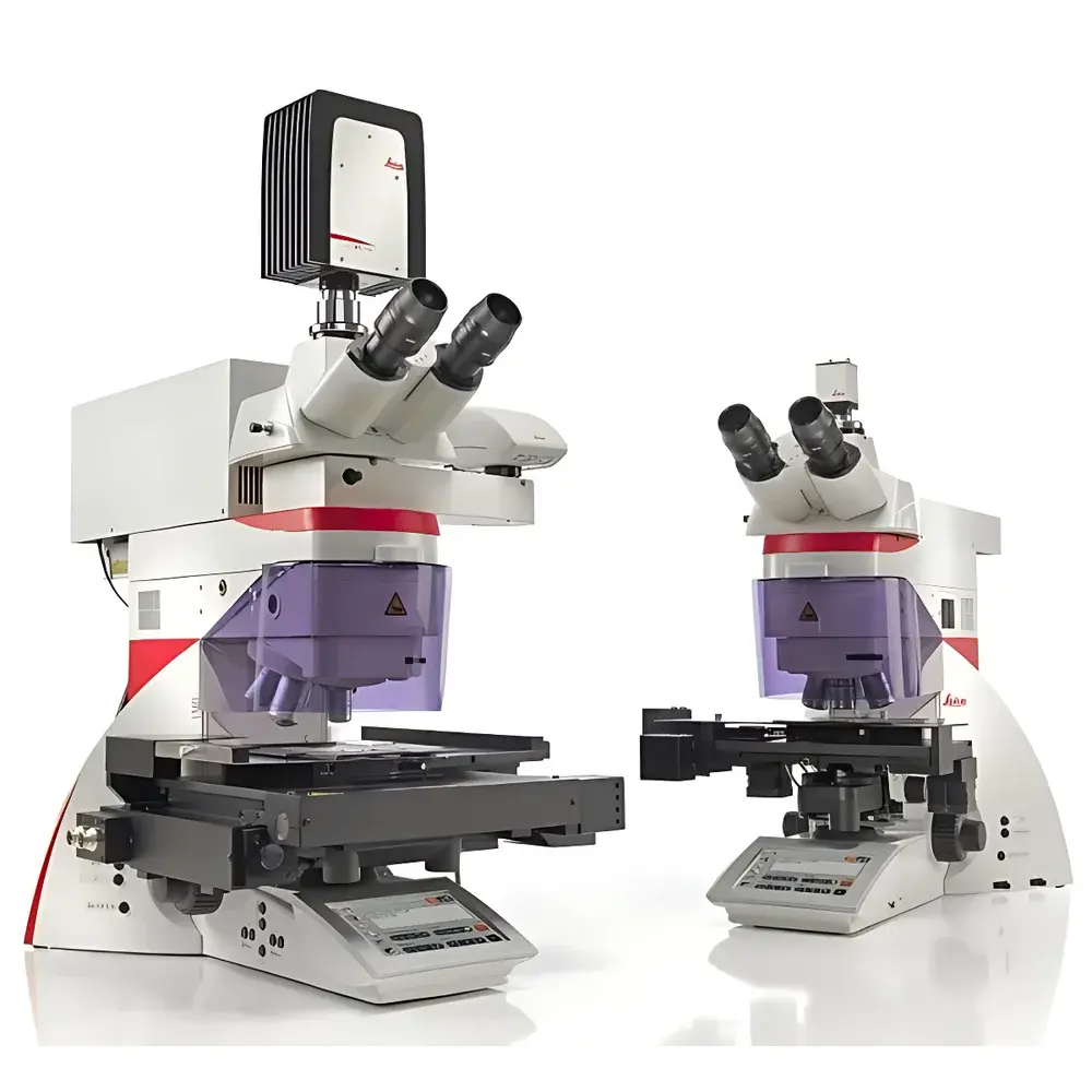
Leica LMD7 Laser Microdissection System
| Brand | Leica |
|---|---|
| Country of Origin | Germany |
| Model | LMD7 |
| Laser Wavelength | 349 nm |
| Pulse Repetition Rate | 10–5000 Hz |
| Pulse Duration | < 4 ns |
| Maximum Pulse Energy | 120 µJ |
| Optical Collection Method | Gravity-based |
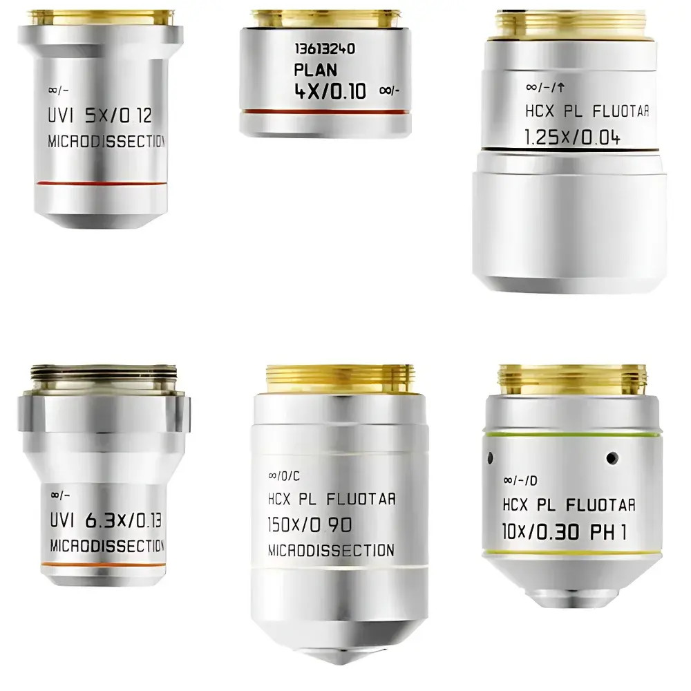
| Compatible Objectives | 5× to 150× dry objectives (SmartCut series) |
| Illumination Options | LED (25,000 h lifetime, constant CCT) or Halogen with CCIC |
| Cutting Modes | "Move-Cut" (direct real-time laser scanning) and "Draw-Scan" (point-ablation without film) |
| Sample Compatibility | FFPE tissues, frozen sections, plant material, chromosomes, bone, teeth, brain, live cells in culture |
| Collection Vessels | Standard 0.2 mL/0.5 mL PCR tubes, multiwell plates, ibidi µ-slides, PEN-membrane slides, PET slides, DIRECTOR slides |
Overview
The Leica LMD7 Laser Microdissection System is a high-precision, upright optical instrument engineered for contact-free, contamination-free isolation of morphologically defined regions from heterogeneous tissue sections or cultured cells. It operates on the principle of ultraviolet (UV) laser ablation—specifically, pulsed UV laser beam steering via high-accuracy galvanometric mirrors and prism-based optical path control—enabling precise spatial targeting without mechanical movement of the specimen stage. Unlike conventional microdissection tools that rely on physical contact or sample translation, the LMD7 moves only the laser focus across the static sample plane, ensuring subcellular resolution, minimal thermal damage, and preservation of molecular integrity. This optical design delivers vertical, perpendicular incisions relative to the tissue surface, critical for downstream genomic, transcriptomic, proteomic, and metabolomic analyses requiring pristine biomolecular input. The system integrates seamlessly into GLP- and GMP-aligned workflows, supporting traceable, auditable sample handling per ISO 15189 and FDA 21 CFR Part 11 requirements when paired with optional audit trail and electronic signature modules.
Key Features
- Gravity-Based Collection Architecture: Eliminates need for adhesive caps or vacuum transfer; dissected regions fall freely under gravity into standard molecular biology receptacles (e.g., 0.2 mL PCR tubes, 96-well plates, ibidi µ-slides), ensuring zero cross-contamination and full compatibility with existing labware.
- High-Flexibility UV Laser Engine: 349 nm wavelength, <4 ns pulse duration, 120 µJ maximum pulse energy, and continuously adjustable repetition rate (10–5000 Hz) enable optimized ablation across diverse sample types—from soft neural tissue to mineralized bone or plant cell walls—without compromising structural fidelity.
- SmartCut Objective Series: Ten dry objective options (5×–150×), all optimized for UV transmission down to 350 nm. The 150× SmartCut objective delivers diffraction-limited resolution at high magnification for chromosome-level dissection; lower-magnification optics support rapid macro-isolation of large tissue domains.
- Dual Illumination Platform: Switchable LED (25,000 h lifetime, stable 5700 K CCT, 90% energy reduction vs. halogen) or halogen illumination with Constant Color Temperature Control (CCIC), preserving color fidelity and enabling reproducible brightfield imaging across long-term experiments.
- Two Native Cutting Paradigms: “Move-Cut” mode enables real-time, freehand laser tracing on PEN-membrane slides for maximal speed and visual feedback; “Draw-Scan” mode supports film-free ablation on standard glass, DIRECTOR, or PET slides—ideal for proteomics/metabolomics where plasticizer interference must be avoided.
Sample Compatibility & Compliance
The LMD7 accommodates a broad spectrum of biological specimens: formalin-fixed paraffin-embedded (FFPE) and cryosections, fresh-frozen tissues, plant sections, cytological preparations, metaphase spreads, decalcified bone, dental enamel, and live adherent cells cultured on ibidi µ-slides or PEN-coated dishes. For live-cell applications, optional climate chamber integration maintains CO2, humidity, and temperature control during dissection. All collection workflows comply with ISO/IEC 17025 analytical validity criteria and support traceability required by CLIA, CAP, and EU IVDR frameworks. When used with PET or DIRECTOR slides, the system meets stringent low-background requirements for mass spectrometry-based proteomics and metabolomics—validated per ASTM E2567 for biomolecular recovery efficiency.
Software & Data Management
LMD Software v7.6 provides a workflow-centric, touch-optimized interface designed for routine laboratory operation. Core functions include panoramic slide mapping, ROI annotation via polygonal or freehand selection, real-time laser parameter adjustment (pulse energy, frequency, aperture, scan speed), and synchronized time-lapse image capture. Optional modules extend capability: Automated Cell Recognition (ACR) uses machine learning–based pattern recognition to identify and isolate cell populations based on morphology or immunofluorescence intensity; Database Manager links sample metadata (patient ID, staining protocol, section thickness) to exported RNA/DNA files for LIMS integration. All software operations generate timestamped, user-attributed audit logs compliant with 21 CFR Part 11 Annex 11 and EU GMP Annex 11 standards.
Applications
- Single-cell and regional transcriptomics from tumor microenvironments or developmental tissue gradients
- Genome-wide methylation profiling of histopathologically defined epithelial vs. stromal compartments
- Proteomic analysis of laser-captured neurons or amyloid plaques using label-free LC-MS/MS
- Metabolite mapping from plant vascular bundles or root nodules via DESI-MS coupling
- Clonal expansion of isolated live cells for functional assays or organoid derivation
- Chromosome microdissection for FISH probe generation or array CGH target enrichment
FAQ
How does gravity-based collection ensure molecular integrity?
Gravity delivery eliminates shear forces, vacuum-induced dehydration, and adhesive residue—preserving native RNA, protein conformation, and post-translational modifications essential for sensitive downstream assays.
Can the LMD7 be used for live-cell dissection under physiological conditions?
Yes—when coupled with an environmental chamber and ibidi µ-slides or PEN-coated dishes, the system supports real-time dissection of viable adherent cells with >95% post-isolation viability confirmed by trypan blue exclusion and colony-forming unit assays.
What slide types are recommended for proteomics applications?
PET slides (low-plasticizer polymer) or DIRECTOR slides (film-free glass) are validated for minimal background in bottom-up proteomics workflows; both exhibit <0.5 fmol/cm² leachable contaminants per ASTM D7823.
Is the LMD7 compatible with existing laboratory informatics systems?
Yes—the software exports annotated TIFF/OME-TIFF images and tabular ROI coordinates (CSV/JSON), and supports HL7/FHIR-compliant metadata exchange with major LIS and ELN platforms via REST API.
How is laser calibration maintained over time?
The system performs automated daily alignment checks using internal reference photodiodes and NIST-traceable power sensors; calibration certificates are generated per ISO/IEC 17025 and archived with each session’s audit log.


