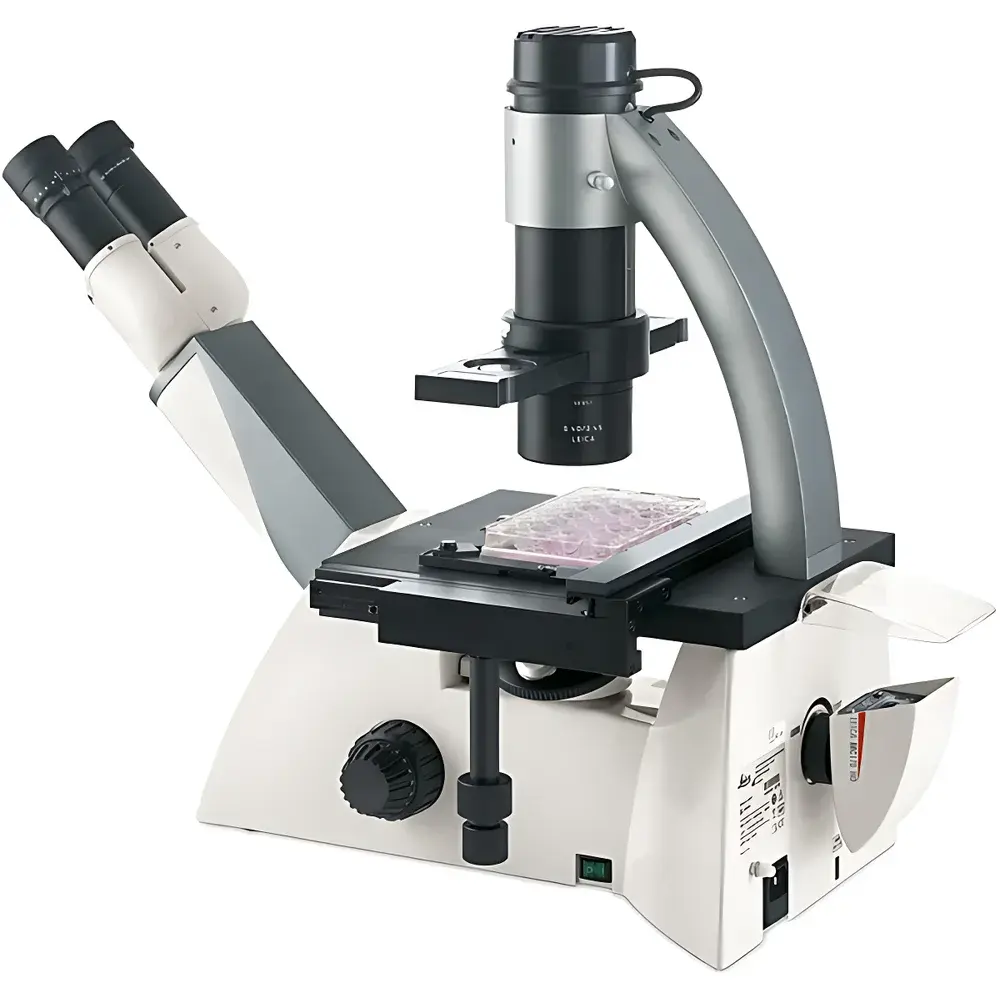
Leica DMi1 Inverted Microscope
| Brand | Leica |
|---|---|
| Origin | Shanghai, China |
| Manufacturer Type | Authorized Distributor |
| Country of Origin Classification | Domestic (China) |
| Model | DMi1 |
| Price Range | USD 7,000 – 14,000 |
| Instrument Category | Inverted Microscope |
| Eyepiece Configuration | Binocular |
Overview
The Leica DMi1 is an entry-level inverted microscope engineered for routine live-cell observation and basic tissue culture monitoring in academic, clinical, and industrial life science laboratories. Designed around the principles of Köhler illumination and phase contrast optics, the DMi1 delivers consistent, high-contrast monochrome imaging without fluorescence capability. Its optical path is optimized for transmitted light techniques—primarily brightfield and phase contrast—making it ideal for unstained or lightly stained adherent and suspension cell cultures grown in standard labware including Petri dishes, multi-well plates, and T-flasks. The system features a fixed-height stage with mechanical X–Y translation, a robust all-metal chassis, and a low center of gravity to minimize vibration-induced image drift during extended observation sessions. Unlike research-grade inverted platforms, the DMi1 omits motorized components, autofocus, and encoded hardware, prioritizing operational simplicity, long-term mechanical stability, and cost-effective maintenance.
Key Features
- Phase contrast compatibility across 10×, 20×, and 40× objectives using a single PH1 annulus—no manual ring exchange required during magnification switching
- Dual working distance capability: S40 condenser (40–50 mm WD) for standard culture vessels; tool-free upgrade path to S80 condenser (80 mm WD) for taller flasks—no reconfiguration of illumination arm needed
- Integrated 5 W LED illumination with constant 5,600 K color temperature across full intensity range; eliminates warm-up time and thermal drift at specimen plane
- Auto-intensity sensor embedded in phase contrast slider adjusts illumination level automatically when switching between brightfield and phase contrast modes
- Energy-efficient design: LED lifetime rated at ≥20 years (based on 40 hours/week usage); 2-hour auto-shutdown function reduces standby power consumption
- Binocular viewing head with ergonomic eyepoint height adjustment and interpupillary distance calibration (55–75 mm)
- Anti-scratch aluminum alloy stage surface with precision-machined mechanical stage controls and engraved scale markers
Sample Compatibility & Compliance
The DMi1 accommodates common in vitro culture formats—including 35 mm, 60 mm, and 100 mm Petri dishes; 6-, 12-, 24-, 48-, and 96-well plates; and T-25, T-75, and T-175 flasks—without requiring custom adapters. Its modular condenser system ensures optimal numerical aperture alignment and Köhler illumination across varying vessel heights. While not certified for GMP or GLP-regulated environments out-of-the-box, the instrument supports traceable documentation workflows when paired with optional LAS software and HDMI-connected display systems. All optical components comply with ISO 10934-1 (Microscopes — Nomenclature of parts) and meet CE marking requirements under EU Directive 2014/30/EU (EMC) and 2014/35/EU (LVD). No IEC 61000-4 immunity testing data is provided for laboratory electromagnetic environments.
Software & Data Management
The optional DMi1 Camera version integrates a 2.5 MP or 5 MP CMOS sensor mounted directly on the rear port—eliminating the need for trinocular head modification or external beam splitters. Image and video capture is controlled via IR remote; output is saved natively to removable SD card (Class 10, up to 128 GB). HDMI output enables real-time projection onto external monitors for collaborative review or teaching. For post-acquisition analysis, the bundled Leica Application Suite (LAS) Core edition provides measurement tools (length, area, angle), annotation overlays, basic histogram analysis, and export in TIFF, JPEG, and BMP formats. LAS Core does not support automated cell counting, Z-stack reconstruction, or regulatory-compliant audit trails per FDA 21 CFR Part 11.
Applications
- Routine daily assessment of mammalian and insect cell confluency in CO₂ incubators and laminar flow hoods
- Monitoring morphology changes during transfection, drug treatment, or serum starvation protocols
- Quality control of primary cell isolations and passage validation prior to cryopreservation
- Documentation of colony formation assays and wound-healing migration experiments
- Instructional microscopy in undergraduate biology and biotechnology training labs
- Pre-screening of samples prior to downstream processing (e.g., FACS sorting, RNA extraction, immunostaining)
FAQ
Is the Leica DMi1 suitable for fluorescence imaging?
No. The DMi1 lacks excitation filters, dichroic mirrors, and high-sensitivity emission detection pathways required for fluorescence microscopy. It is strictly a transmitted-light platform.
Can the DMi1 be upgraded to support DIC or Hoffman modulation contrast?
No. The optical train and condenser design are limited to brightfield and phase contrast. DIC and other contrast enhancement methods require dedicated prism sets and specialized condensers not compatible with the DMi1 architecture.
Does the DMi1 include objective lenses in the base configuration?
Yes. Standard configurations include Plan Achromat 10×, 20×, and 40× phase contrast objectives with spring-loaded correction collars and PH1 alignment capability.
What is the maximum usable magnification with the included eyepieces?
With 10× wide-field eyepieces and the 40× objective, total magnification is 400×—sufficient for subcellular feature resolution in cultured cells but not for ultrastructural detail.
Is service and calibration support available outside mainland China?
Yes. Leica Microsystems maintains authorized service centers in over 40 countries; calibration certificates and preventive maintenance contracts are available upon request through local distributors.


