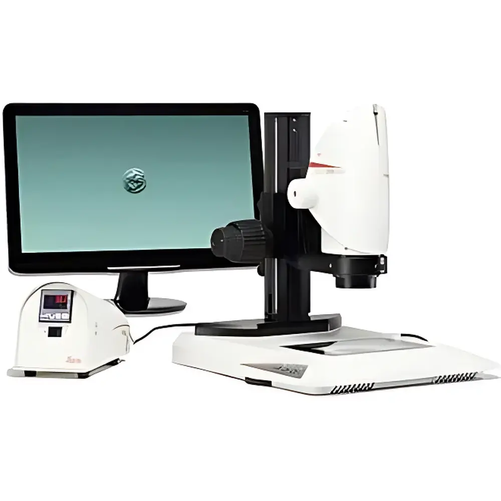
Leica DMS1000 B Digital Stereo Microscope System
| Brand | Leica |
|---|---|
| Origin | Germany |
| Model | DMS1000 B |
| Optical Magnification | Up to 300× |
| Camera Resolution | 5 MP |
| Video Frame Rate | 30 fps (Full HD) |
| Illumination | Integrated Transmitted Light Base (TL3000 ST or TL5000 ergo) |
| Standalone Operation | Yes (SD card storage, IR remote control) |
| Ergonomic Design | Fully eye-piece-free, monitor-based viewing |
| Calibration | Encoded zoom with auto-scaling annotation |
Overview
The Leica DMS1000 B is a fully integrated, standalone digital stereo microscope system engineered for precision-critical life science applications—particularly those requiring strict environmental control and operator safety. Unlike conventional stereomicroscopes relying on ocular observation, the DMS1000 B employs a high-sensitivity CMOS imaging sensor coupled with Leica’s apochromatic optical pathway to deliver real-time, distortion-corrected stereoscopic visualization at magnifications up to 300×. Its core measurement principle is based on dual-path optical beam splitting and digital image fusion, enabling true depth perception without binocular strain. Designed explicitly for use inside laminar flow hoods and cleanroom environments—including IVF laboratories, stem cell culture facilities, and embryology suites—the system eliminates the need for eyepieces, thereby removing direct operator contact with optical ports and minimizing contamination risk per ISO 14644-1 Class 5 (or equivalent) cleanroom protocols.
Key Features
- Fully self-contained architecture: Integrated 5 MP CMOS camera, LED-transmitted illumination base (interchangeable between TL3000 ST and TL5000 ergo models), and embedded image processing unit—no external PC required.
- Real-time Full HD imaging: Sustained 30 fps output at 1920 × 1080 resolution ensures fluid motion capture during micromanipulation tasks such as oocyte handling, blastocyst biopsy, or microinjection.
- Encoded zoom optics: Motorized, calibrated zoom system automatically annotates scale bars synchronized to magnification settings—supporting traceable documentation compliant with GLP and ISO/IEC 17025 requirements.
- Infrared remote operation: Intuitive one-button interface enables rapid switching between still capture, video recording (up to several seconds), and live viewing modes—optimized for glove-compatible workflows.
- Ergonomic workflow integration: Monitor-based visualization eliminates inter-user ocular alignment adjustments; adjustable display height and angle accommodate diverse anthropometric profiles without compromising posture integrity.
- Onboard storage: Internal SD card slot supports lossless image export in TIFF and JPEG formats, plus MP4 video files—facilitating immediate review and audit-ready data retention.
Sample Compatibility & Compliance
The DMS1000 B accommodates transparent, semi-transparent, and low-contrast biological specimens—including zygotes, cleavage-stage embryos, blastocysts, and dissociated stem cell colonies—without staining or fixation. Its transmitted-light base provides uniform Köhler illumination across variable specimen thicknesses (0.1–5 mm), while optional contrast enhancement modules (e.g., polarizing filters, oblique lighting adapters) extend utility to non-biological samples such as microelectronics or polymer films. The system complies with IEC 61000-6-3 (EMC emissions), IEC 61000-6-2 (immunity), and UL/CSA 61010-1 for laboratory equipment safety. Its sealed optical housing meets IP20 ingress protection standards, ensuring operational reliability within ISO Class 5 cleanrooms when installed per manufacturer-specified mounting guidelines.
Software & Data Management
While the DMS1000 B operates independently, optional Leica Application Suite (LAS X) software enables advanced post-acquisition analysis—including multi-focus stacking, region-of-interest (ROI) measurement, annotation layers, and batch metadata tagging. All onboard images and videos embed EXIF-compliant metadata (timestamp, magnification, illumination intensity, focus position). Audit trail functionality—when used with LAS X in networked configurations—supports FDA 21 CFR Part 11 compliance through electronic signatures, user access logs, and immutable record archiving. Export formats include DICOM-SR for PACS integration and CSV for LIMS synchronization.
Applications
- In vitro fertilization (IVF) and assisted reproductive technology (ART): Embryo grading, intracytoplasmic sperm injection (ICSI), assisted hatching, and blastocyst vitrification monitoring.
- Stem cell research: Colony morphology assessment, passaging verification, and differentiation stage documentation under sterile conditions.
- Cell therapy manufacturing: Release testing of autologous/allogeneic cell products per USP sterility requirements and EU Annex 1 GMP guidelines.
- Developmental biology: Time-lapse imaging of early embryogenesis in model organisms (e.g., zebrafish, mouse), where minimal phototoxicity and stable focus are critical.
- Microsurgery training: High-fidelity simulation of microvascular anastomosis and nerve repair techniques using ex vivo tissue models.
FAQ
Does the DMS1000 B require a computer for basic operation?
No. All core imaging functions—including capture, playback, zoom, and illumination control—are managed via the built-in interface and IR remote. A computer is only necessary for advanced analysis using optional LAS X software.
Can the system be validated for GxP-regulated environments?
Yes. With documented IQ/OQ protocols, encoded zoom calibration certificates, and optional 21 CFR Part 11–enabled LAS X deployment, the DMS1000 B supports full validation under GMP, GLP, and CLIA frameworks.
What is the maximum working distance achievable at highest magnification?
At 300× total magnification, the working distance is approximately 42 mm—sufficient to accommodate standard Petri dishes, culture dishes, and micromanipulation tools inside most Class II biosafety cabinets.
Is the TL5000 ergo base compatible with temperature-controlled stages?
Yes. The TL5000 ergo features a modular design with standardized mounting interfaces, allowing seamless integration with third-party heated/cooled stages rated for ±0.1°C stability—essential for time-lapse embryo culture.
How is focus stability maintained during extended imaging sessions?
The system incorporates passive thermal compensation in its objective lens group and uses a fixed-focus optical path architecture—eliminating mechanical drift associated with motorized focus mechanisms. Focus consistency is verified per ISO 9345-2 over 8-hour continuous operation.


