DLAB DL-SUB03+ Multi-Functional Blue Light Transilluminator Electrophoresis Tank
| Brand | DLAB |
|---|---|
| Origin | Beijing, China |
| Manufacturer Type | Direct Manufacturer |
| Country of Origin | China |
| Model | DL-SUB03+ |
| Pricing | Upon Request |
| Gel Tray Dimensions (W×L) | 13×20 cm and 13×15 cm |
| Comb Options | 1.0 mm (14-, 18-, 26-well), 1.5 mm (18-well) |
| Buffer Capacity | 800 mL |
| Electrode Material | 99.99% Pure Platinum Alloy, Ø0.2 mm |
| Tank Dimensions (L×W×H) | 340×170×130 mm |
| Weight | 1.2 kg |
| Construction | High-Impact Polycarbonate Injection-Molded Body |
| Auto Shut-off | 5 min |
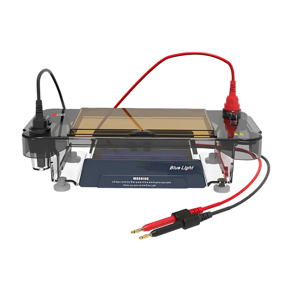
Overview
The DLAB DL-SUB03+ is a purpose-engineered horizontal electrophoresis system designed for high-throughput nucleic acid separation under blue-light excitation. Unlike conventional UV-based systems, it integrates a uniform matrix of high-efficiency blue LEDs (peak emission ~470 nm) beneath the gel tray, enabling real-time visualization of DNA bands during electrophoresis when used with blue-light–compatible nucleic acid stains (e.g., SYBR Safe, GelGreen, or BlueView dyes). This eliminates the need for post-run UV transillumination—reducing photodamage to DNA, minimizing mutagenic exposure risk, and supporting safer laboratory workflows compliant with occupational health guidelines (OSHA 1910.132, IEC 62471). The system operates on standard DC power input and is optimized for agarose and polyacrylamide gel electrophoresis (AGE/PAGE) applications in molecular biology, genotyping, and quality control laboratories.
Key Features
- Integrated blue LED transilluminator with uniform irradiance distribution across both gel formats (13×15 cm and 13×20 cm), ensuring consistent band visibility without hotspots or shadowing.
- Patented flip-top anti-blue-light shield constructed from optical-grade polycarbonate with >99.9% blue light filtration—enabling unaided visual inspection of resolved bands under ambient lighting conditions.
- Programmable 5-minute auto-shutoff circuitry protects LED longevity and reduces thermal load during extended use; manual override allows continuous operation when required.
- Modular electrode assembly with detachable platinum alloy (99.99% purity) wire electrodes (Ø0.2 mm), facilitating rapid cleaning, replacement, and maintenance without disassembling the tank body.
- Precision-machined leveling feet at the base allow fine adjustment of horizontal alignment—critical for uniform electric field distribution and reproducible migration patterns.
- One-piece injection-molded polycarbonate chassis provides structural rigidity, chemical resistance to common electrophoresis buffers (TAE, TBE, SB), and zero leakage integrity under static and dynamic buffer loads.
- Dual-format compatibility supported by interchangeable gel trays and comb holders—optimized for routine analysis (13×15 cm) and high-density screening (13×20 cm, up to 200 samples per run).
Sample Compatibility & Compliance
The DL-SUB03+ is validated for use with standard agarose (0.7–3.0%) and native/polyacrylamide gels (3–20%). Its design accommodates both low-voltage (≤150 V) and medium-voltage (up to 250 V) electrophoretic protocols, delivering stable current output within ±2% variation over 90-minute runs. The system conforms to ISO 13485–aligned manufacturing practices and meets electrical safety requirements per IEC 61010-1:2010 (Equipment for Laboratory Use). While not a medical device, its materials comply with EU REACH Annex XVII restrictions and are free of SVHC substances above threshold limits. No regulatory submission (e.g., FDA 510(k)) applies, as it falls outside diagnostic instrument classification per 21 CFR Part 866.
Software & Data Management
As a standalone electrophoresis cell, the DL-SUB03+ does not incorporate embedded firmware or digital connectivity. It is fully compatible with third-party imaging systems (e.g., Azure Biosystems cSeries, Bio-Rad ChemiDoc MP) via standard gel documentation workflows. All physical controls—including electrode polarity switching and buffer level monitoring—are manually operated, aligning with GLP principles requiring audit-trail transparency and operator accountability. Documentation of gel run parameters (voltage, time, buffer composition) is maintained externally in lab notebooks or LIMS platforms compliant with 21 CFR Part 11 when paired with electronic signature-capable software.
Applications
- Routine DNA fragment analysis in academic and clinical research labs, including PCR product verification, restriction digest profiling, and plasmid QC.
- High-throughput genotyping workflows requiring parallel sample processing—leveraging dual-comb configurations and 200-sample capacity per run.
- Educational electrophoresis demonstrations where student safety and real-time observation enhance pedagogical outcomes.
- Quality assurance testing of oligonucleotide synthesis batches, where blue-light visualization preserves sample integrity for downstream sequencing or cloning.
- Field-deployable molecular diagnostics support (when paired with portable power supplies), particularly in resource-limited settings where UV infrastructure is unavailable or hazardous.
FAQ
Is the DL-SUB03+ compatible with ethidium bromide (EtBr)?
No—EtBr requires UV excitation (~302 nm) and exhibits negligible fluorescence under blue light. Use only blue-light–optimized dyes such as SYBR Safe, GelRed (blue variant), or LuminoBlue.
Can the blue LED module be replaced independently?
Yes—the LED array is mounted on a serviceable PCB subassembly secured with four M2.5 screws; replacement kits are available directly from DLAB Technical Support.
What buffer volumes are recommended for each gel size?
For the 13×15 cm tray: 450–600 mL; for the 13×20 cm tray: 650–800 mL. Maintain minimum buffer depth of 3 mm above the gel surface to ensure uniform current density.
Does the system include a built-in power supply?
No—it is a passive electrophoresis cell requiring an external DC power supply (0–300 V, 0–500 mA) with adjustable voltage/current limiting.
How is electrode polarity reversed for bidirectional electrophoresis?
By physically swapping the red/black alligator clip connections to the platinum electrodes—no internal switching mechanism is provided, preserving signal integrity and reducing failure points.
Related Products


