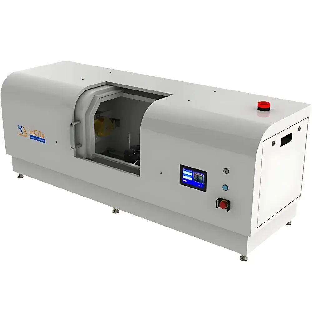
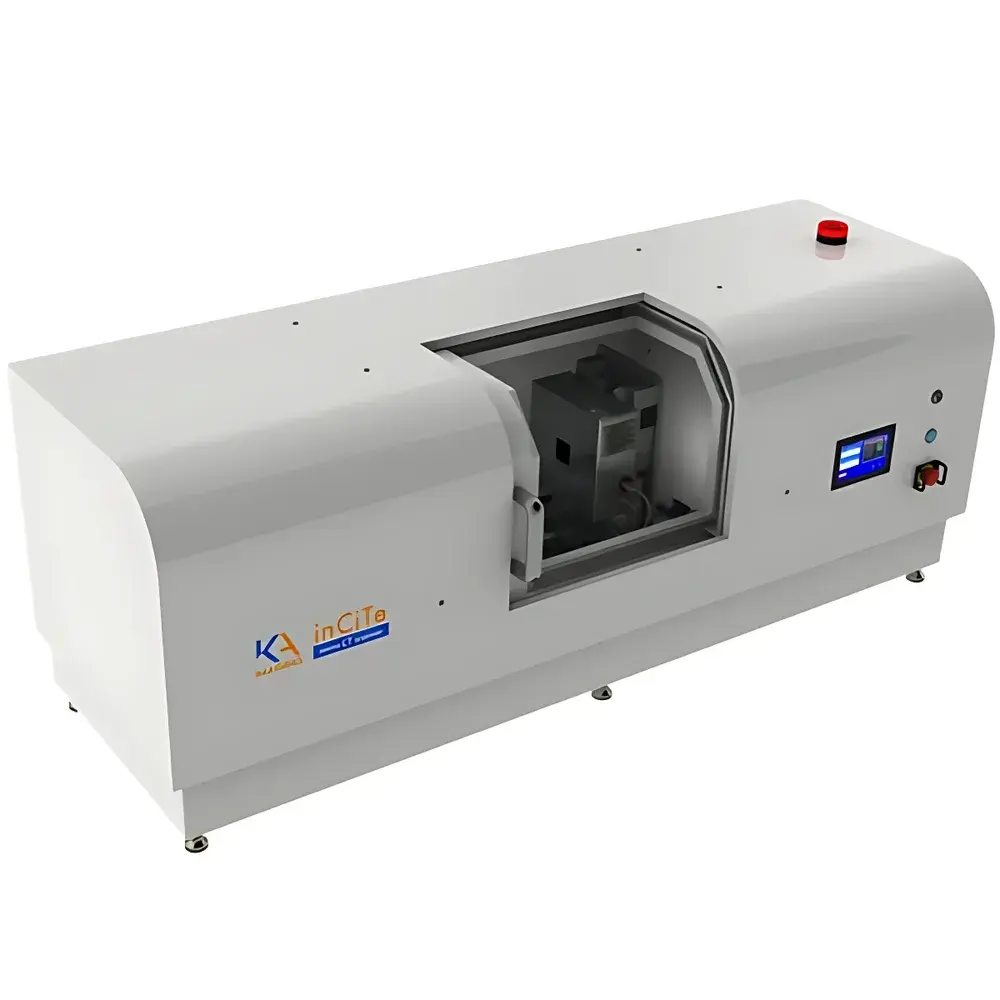
KA Imaging inCiTe 3D Desktop Phase-Contrast Micro-CT System with 2 μm Focal Spot
| Brand | KA Imaging |
|---|---|
| Origin | Canada |
| Model | inCiTe 3D |
| Detector Type | High-Resolution CMOS-Based Direct-Conversion a-Se (BrillianSe) Sensor |
| Scan Mode | Rotation-Only (RO) |
| Spatial Resolution | Up to 5.6 μm (0.1 MTF at 90 cycles/mm) |
| X-ray Energy Range | 40–110 kV |
| Field of View | 32 mm × 32 mm (direct detection) |
| Maximum Sample Dimensions | Ø25 mm × 100 mm (diameter × height/length) |
| System Footprint | 1500 mm × 580 mm × 500 mm (L × W × H) |
| Pixel Pitch | 8 μm |
| Detector Resolution | 4096 × 4096 (16.8 MP) |
| Effective DQE | High across 40–120 keV |
| Phase-Contrast Mechanism | Propagation-Based (Grating-Free, In-Line) |
| Minimum Focal Spot Size | 2 μm |
Overview
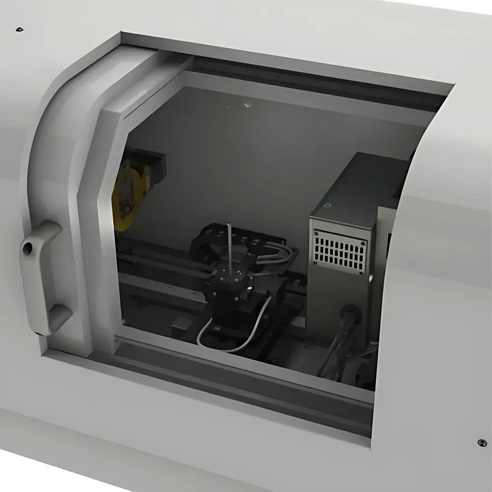
The KA Imaging inCiTe 3D Desktop Phase-Contrast Micro-CT System is a high-resolution, laboratory-scale X-ray microscopy platform engineered for quantitative 3D non-destructive evaluation of low-absorbing and heterogeneous materials. Unlike conventional absorption-based micro-CT systems, the inCiTe 3D leverages propagation-based phase-contrast imaging (PCI), a physically distinct contrast mechanism that exploits X-ray wavefront phase shifts rather than attenuation differences. This enables orders-of-magnitude improvement in soft-tissue and low-Z material contrast—particularly critical for biological specimens, polymers, composites, pharmaceuticals, and additively manufactured components where traditional CT yields poor signal-to-noise ratios. At its core lies KA Imaging’s proprietary BrillianSe detector: a monolithic, direct-conversion amorphous selenium (a-Se) / CMOS hybrid sensor with 8 µm pixel pitch, 16.8 megapixel resolution, and high detective quantum efficiency (DQE) up to 120 keV. Coupled with a microfocus X-ray source featuring a 2 µm focal spot, the system achieves spatial resolution down to 5.6 µm (0.1 MTF at 90 cycles/mm) under optimized geometry—making it suitable for sub-cellular morphology analysis, pore network characterization, and micro-defect detection without staining, sectioning, or contrast agents.
Key Features
- Propagated-phase contrast imaging (grating-free, in-line geometry) for enhanced edge enhancement and soft-tissue visibility
- BrillianSe direct-conversion a-Se/CMOS detector: 4096 × 4096 pixels, 8 µm pitch, high DQE across 40–120 keV range
- Microfocus X-ray source with 2 µm minimum focal spot size for high geometric magnification and resolution scalability
- Rotation-only (RO) scanning architecture ensuring mechanical stability, repeatability, and minimal vibration-induced artifacts
- Large active field of view: 32 mm × 32 mm—enabling high-throughput imaging of multi-sample trays or extended specimens
- Compact desktop footprint (1500 × 580 × 500 mm) with integrated shielding, eliminating need for dedicated vault infrastructure
- Zero-sample-prep workflow: compatible with native, hydrated, or uncoated specimens—including live small-animal tissues and polymer foams
Sample Compatibility & Compliance
The inCiTe 3D accommodates a broad spectrum of sample types relevant to industrial QA/QC and preclinical research: metallic implants (e.g., titanium lattice structures), soft biological tissues (murine knee joints, renal calculi), pharmaceutical dosage forms (capsules, tablets), electronic assemblies (PCBs, solder joints), fiber-reinforced composites (aramid, carbon, flax), and construction materials (lightweight aggregate concrete, aerogels). Its phase-sensitive contrast mechanism satisfies requirements outlined in ASTM E2737 (Standard Practice for Digital Radiographic Testing), ISO 15708-2 (Non-destructive testing — Qualification of computed tomography systems), and supports GLP-compliant workflows through audit-trail-enabled software logging. While not FDA-cleared for human diagnostics, the system meets IEC 62464-1 radiation safety standards and complies with Canadian Radiation Emitting Devices Regulations (REDR) and EU Directive 2013/59/Euratom for occupational exposure control.
Software & Data Management
Acquisition and reconstruction are managed via KA Imaging’s inCiTe Control Suite—a Windows-based application with intuitive GUI designed for both novice operators and advanced users. The suite provides real-time projection preview, automated geometry calibration (center-of-rotation, beam hardening correction), and one-click alignment tools. Reconstruction employs GPU-accelerated filtered back-projection (FBP) and iterative algorithms (SART, SIRT) supporting ring artifact suppression, beam-hardening correction, and phase-retrieval post-processing. Output formats include DICOM, TIFF stacks, and HDF5—ensuring compatibility with third-party visualization platforms (Avizo, Dragonfly, VGStudio MAX) and AI training pipelines. All acquisition parameters, user actions, and timestamped metadata are recorded in encrypted logs compliant with 21 CFR Part 11 principles for electronic records and signatures—supporting traceability in regulated environments.
Applications
- Microstructural characterization of porous media, foams, and biomaterial scaffolds
- Reverse engineering of legacy parts via high-fidelity surface mesh extraction (STL/OBJ export)
- Validation of multiphysics simulation models (e.g., fluid flow in rock cores, thermal stress in AM lattices)
- NDT inspection per ASME BPVC Section V and ISO 17636-2 for internal voids, delaminations, and residual powder in additively manufactured components
- GD&T analysis including form, position, and profile tolerances against CAD reference models
- In-process monitoring of sintering, curing, or hydration dynamics using time-resolved 4D micro-CT
- Root-soil interaction studies, seed germination tracking, and plant vascular network mapping in agricultural research
FAQ
Does the inCiTe 3D require synchrotron radiation to achieve phase contrast?
No. It operates with a laboratory-based microfocus X-ray tube and relies on free-space propagation (in-line geometry), eliminating the need for gratings or synchrotron sources.
Can the system perform quantitative density calibration?
Yes—via dual-energy acquisition and reference phantoms calibrated against known attenuation coefficients, enabling approximate electron density mapping.
Is the BrillianSe detector sensitive to beam hardening artifacts?
The detector’s high DQE and linear response across 40–110 kV reduce spectral dependency; however, standard beam-hardening correction algorithms are applied during reconstruction.
What is the typical scan time for a full 360° acquisition at 5 µm voxel resolution?
Scan duration depends on sample attenuation and desired SNR, but typical acquisition ranges from 15 to 45 minutes for medium-density samples (e.g., polymer composites, bone tissue) using automatic exposure optimization.
Does the system support time-lapse (4D) imaging?
Yes—through programmable sequential scans with automated stage repositioning and synchronized trigger logic for dynamic process monitoring.


