
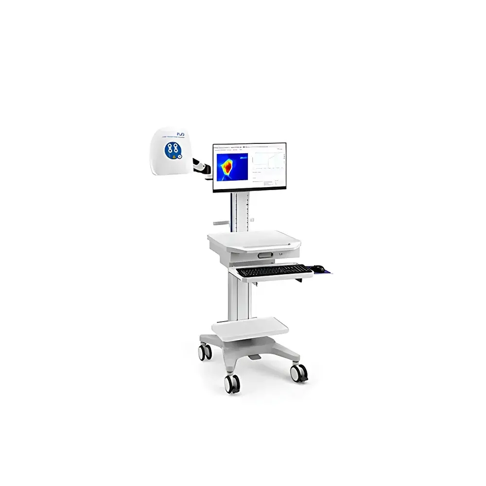
RWD RFSLI ZW Laser Speckle Contrast Imager
| Brand | RWD |
|---|---|
| Origin | Shenzhen, China |
| Manufacturer Type | Original Equipment Manufacturer (OEM) |
| Product Category | Domestic |
| Model | RFSLI ZW |
| Instrument Type | Optical Imaging System |
| Maximum Scan Resolution | 2048 × 2048 pixels |
| Frame Rate | 120 FPS |
| Field of View | 90 mm × 90 mm |
| Simultaneous Sample Capacity | 4 |
| Spatial Resolution | 3.9 µm/pixel |
| Laser Power Stability | <1% RMS fluctuation |
| Minimum Perfusion Unit (PU) Resolution | 0.001 PU |
| Optical Zoom | 12× |
| Data Acquisition Mode | Real-time dynamic contrast imaging |
Overview
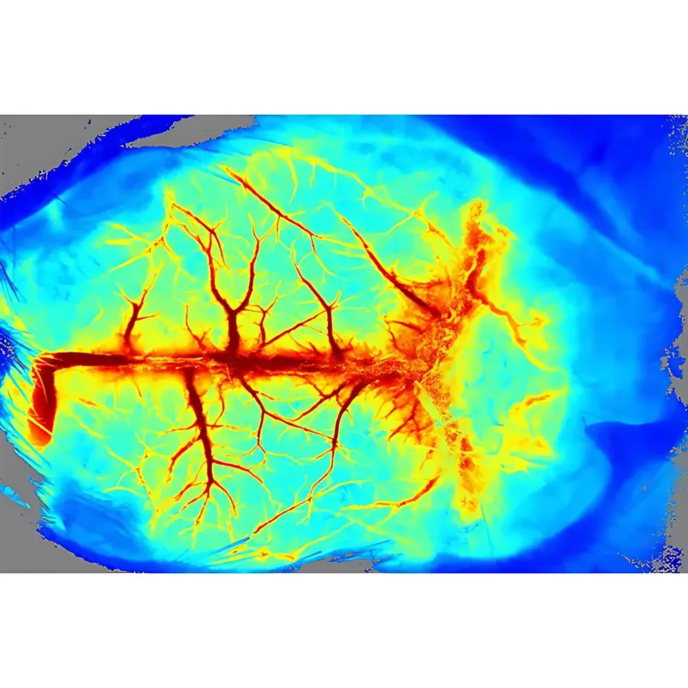
The RWD RFSLI ZW Laser Speckle Contrast Imager is a high-performance, non-contact optical imaging system engineered for quantitative, real-time assessment of microvascular perfusion in small animal models. Based on Laser Speckle Contrast Imaging (LSCI) principles, the system exploits the temporal blurring of laser speckle patterns generated by moving red blood cells to compute relative blood flow velocity and spatial perfusion distribution without exogenous contrast agents. Unlike Doppler-based or fluorescent methods, LSCI provides full-field, label-free, and inherently motion-tolerant hemodynamic mapping—making it ideal for longitudinal studies where physiological integrity must be preserved. The RFSLI ZW delivers sub-cellular spatial resolution (3.9 µm/pixel) across a wide 90 mm × 90 mm field of view, enabling simultaneous monitoring of cortical, spinal, renal, mesenteric, dermal, and tumor-associated vasculature under native physiological conditions.
Key Features
- Full-Field High-Resolution Imaging: Equipped with a scientific-grade CMOS sensor and 12× motorized optical zoom lens, the system supports up to 2048 × 2048 pixel acquisition at native resolution, ensuring precise localization of capillary-level flow changes across heterogeneous tissue domains.
- Ultra-High Temporal Sampling: Capable of sustained 120 FPS acquisition with hardware-triggered synchronization, the RFSLI ZW captures rapid hemodynamic transients—including cortical spreading depression (CSD), ischemia-reperfusion dynamics, and acute vasoactive responses—with minimal motion artifact.
- Quantitative Perfusion Stability: Engineered for longitudinal reproducibility, the system maintains laser power stability below 1% RMS over multi-hour sessions and delivers perfusion quantification with a resolution of 0.001 Perfusion Units (PU), validated against calibrated microsphere and Doppler flow probe benchmarks.
- Multi-Sample Parallel Monitoring: Supports concurrent imaging of up to four independent biological specimens—enabling controlled comparative studies (e.g., sham vs. MCAO, treated vs. vehicle, contralateral limb pairs) within a single acquisition session.
- Modular Software Architecture: Analysis software operates independently of acquisition hardware; data files are saved in open-format HDF5 containers, permitting offline processing on standard Windows workstations without licensing restrictions or node-based activation.
Sample Compatibility & Compliance
The RFSLI ZW is optimized for in vivo use in murine, rat, rabbit, and avian models—including transgenic, immunocompromised, and surgically prepared preparations. It accommodates standard stereotaxic frames, thermal regulation platforms, and gas anesthesia interfaces. All optical components comply with IEC 60825-1:2014 Class 3B laser safety standards, with integrated interlocks and emission indicators. Data acquisition workflows support GLP-compliant metadata tagging (animal ID, time stamp, anesthesia status, physiological parameters) and optional audit trail logging aligned with FDA 21 CFR Part 11 requirements when deployed in regulated preclinical research environments.
Software & Data Management
The bundled RWD LSCI Analysis Suite provides end-to-end processing—from raw speckle contrast computation (using spatiotemporal variance algorithms per ISO/TR 20417:2021 guidance) to region-of-interest (ROI) definition, time-series normalization, and statistical parametric mapping. Users may define custom ROIs across anatomical landmarks (e.g., ischemic penumbra, tumor margin, CAM vascular plexus) and export absolute perfusion values, relative change metrics (%ΔPU), and kinetic parameters (time-to-peak, washout half-life). Batch processing pipelines support automated alignment, motion correction, and group-level statistical comparison (ANOVA, paired t-test) with export to MATLAB, Python (NumPy/Pandas), and Prism-compatible CSV formats.
Applications
- Stroke Research: Validation of middle cerebral artery occlusion (MCAO) success, real-time tracking of collateral recruitment, and quantitative evaluation of neuroprotective or reperfusion therapies across ischemia–reperfusion–recovery timelines.
- Spinal Cord Injury: Dynamic mapping of post-traumatic hypoperfusion and reperfusion injury progression following controlled impactor models, correlating flow deficits with histopathological outcomes.
- Tumor Angiogenesis: Longitudinal quantification of microvascular density and functional perfusion in subcutaneous xenografts (e.g., 4T1, U87MG) during anti-angiogenic or immunomodulatory treatment regimens.
- Renal & Mesenteric Microcirculation: Assessment of sepsis-induced capillary dropout, drug-induced vasomotion, and recovery kinetics in endotoxemia or ischemia-reperfusion models—without nephrectomy or surgical exposure artifacts.
- Dermatological & Translational Studies: Objective evaluation of topical agent efficacy (e.g., vasodilators, anti-inflammatories), wound healing progression, and acupuncture-induced microvascular modulation via standardized ROI-based perfusion analytics.
FAQ
What is the minimum detectable perfusion change the RFSLI ZW can resolve?
The system achieves a perfusion unit (PU) resolution of 0.001 PU under optimal signal-to-noise conditions, corresponding to ~0.5% relative change in capillary flow velocity for typical cortical tissue.
Can the RFSLI ZW be integrated with electrophysiology or behavioral rigs?
Yes—the system supports TTL and analog trigger inputs/outputs for hardware synchronization with EEG amplifiers, optogenetic stimulators, or treadmill systems, enabling multimodal correlation of hemodynamics with neural or motor activity.
Is calibration required before each experiment?
No routine recalibration is needed; factory calibration is traceable to NIST-standard photometric references, and laser stability is continuously monitored via internal photodiode feedback.
Does the software support automated vessel segmentation?
The analysis suite includes semi-automated vessel enhancement filters (Frangi-based) and centerline extraction tools, but manual ROI delineation remains recommended for high-precision microvascular quantification in complex topographies.
How is data integrity ensured during long-term acquisitions?
All acquisitions generate timestamped, checksum-verified HDF5 files with embedded acquisition metadata (exposure time, gain, laser intensity, ambient temperature), supporting FAIR (Findable, Accessible, Interoperable, Reusable) data management principles.


