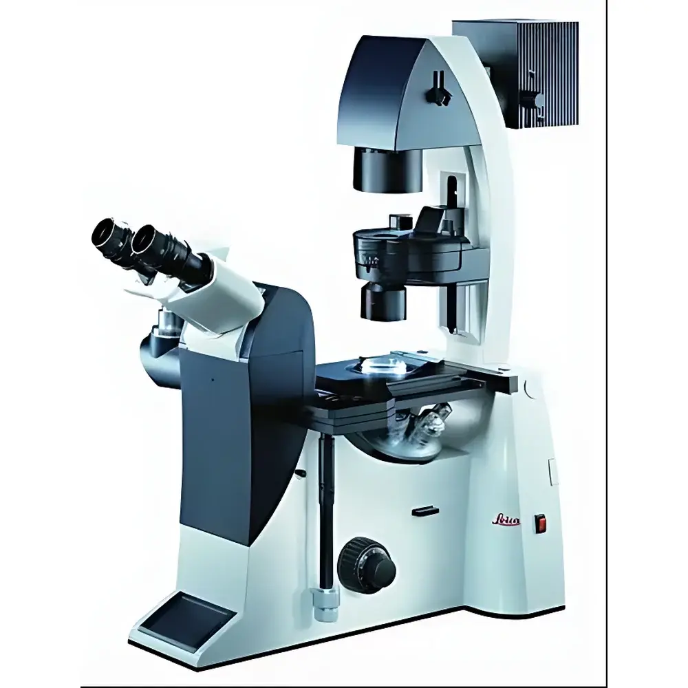
Leica DMI3000 Manual Inverted Microscope
| Brand | Leica |
|---|---|
| Origin | Germany |
| Model | DMI3000 |
| Optical System | Infinity-corrected, Plan Achromatic to Apochromatic Long Working Distance Objectives |
| Objective Turret | 6-position |
| Built-in Zoom | 1.5× |
| Illumination | 12 V / 100 W Halogen |
| Compatibility | Brightfield, Darkfield, Phase Contrast (IPH), Relief Contrast (IMC), DIC, Polarization, Fluorescence, Live-cell Imaging |
| Condenser Options | Universal condenser with interchangeable apertures (1 mm, 23 mm, 28 mm, 40 mm, 70 mm) |
| Software Integration | Compatible with Leica Application Suite (LAS) and LAS X platforms |
| Regulatory Compliance | Designed for GLP/GMP-aligned laboratory environments |
Overview
The Leica DMI3000 Manual Inverted Microscope is a high-precision, research-grade optical platform engineered for demanding life science applications requiring long-term stability, optical fidelity, and modular adaptability. Designed around an infinity-corrected optical pathway, the system delivers consistent resolution and contrast across a full spectrum of transmitted-light contrast techniques—including brightfield, darkfield, differential interference contrast (DIC), polarization, and phase-based modalities—without compromising fluorescence performance. Its inverted configuration positions the objective lenses beneath the specimen stage, enabling direct access to live samples in standard culture vessels (e.g., Petri dishes, flasks, multi-well plates), making it especially suited for time-lapse imaging of adherent and suspension cells, microinjection workflows (e.g., ICSI), and long-duration physiological assays.
Key Features
- Infinity-corrected optical architecture supporting Plan Achromatic through Apochromatic long working distance (LWD) objectives—optimized for thick specimens and cover-glass–compatible cell culture formats.
- 6-position manual objective turret with integrated 1.5× mechanical zoom, allowing rapid magnification adjustment without refocusing or objective switching.
- Dual external contrast modules: IPH (Integrated Phase Contrast Housing) and IMC (Integrated Modulation Contrast) mounted outside the objective path—ensuring zero fluorescence signal loss while enabling seamless transition between phase contrast, relief contrast, and epifluorescence using the same set of fluorescence-optimized objectives.
- Modular illumination design centered on a stable 12 V / 100 W halogen source, with field and aperture diaphragm controls calibrated for Köhler illumination across all contrast modes.
- Universal condenser system with five interchangeable aperture stops (1 mm, 23 mm, 28 mm, 40 mm, 70 mm), facilitating precise numerical aperture matching for optimal resolution and depth discrimination in both routine and advanced imaging protocols.
- Robust mechanical stage options—including manual, motorized, and encoded variants—with compatibility for environmental chambers (CO₂, temperature, humidity control) and micromanipulator integration for microinjection and patch-clamp setups.
Sample Compatibility & Compliance
The DMI3000 accommodates standard and custom sample carriers used in modern cell biology laboratories: glass-bottom dishes (e.g., MatTek, Ibidi), chambered coverslips, multi-well plates (6–96-well), and specialized microfluidic devices. Its long working distance objectives (up to 10.6 mm for 20× LWD) ensure unobstructed access to samples under physiological conditions. The system conforms to ISO 10993 biocompatibility guidelines for materials in contact with biological specimens and meets CE marking requirements for in vitro diagnostic (IVD) support equipment. When operated with Leica LAS X software in validated configurations, the platform supports audit trails, electronic signatures, and role-based user permissions aligned with FDA 21 CFR Part 11 and EU Annex 11 regulatory expectations for data integrity in GLP and GMP environments.
Software & Data Management
The DMI3000 integrates natively with Leica Application Suite (LAS) and LAS X software ecosystems. LAS X enables synchronized hardware control (shutters, filter wheels, focus motors, stage positioning), multi-channel fluorescence acquisition, Z-stack reconstruction, time-series registration, and quantitative intensity profiling. All acquired image metadata—including objective ID, exposure parameters, contrast mode selection, and stage coordinates—are embedded in TIFF and Leica’s native .lif format. Optional modules provide compliance-ready features: electronic signature workflows, change history logging, and secure database export to PACS or LIMS systems via DICOM or HL7 interfaces.
Applications
- Long-term live-cell imaging of primary cultures, stem cell differentiation, and organoid development under controlled environmental conditions.
- Clinical embryology workflows including intracytoplasmic sperm injection (ICSI), blastocyst assessment, and time-lapse morphokinetic analysis.
- Transfection efficiency monitoring, reporter gene expression tracking, and co-localization studies using multi-label fluorescence.
- High-content screening (HCS) preparation and validation—particularly where manual intervention, low phototoxicity, and optical consistency are critical.
- Teaching and core facility use: configured with shared-view optics or HDMI output for classroom demonstration and collaborative microscopy sessions.
FAQ
Can the DMI3000 be upgraded to motorized focusing or automated stage control?
Yes—Leica offers factory-installed motorized Z-drive and encoded XY-stage options compatible with LAS X automation scripting.
Is fluorescence sensitivity compromised when using phase contrast or DIC modules?
No—the IPH and IMC modules are positioned externally to the objective light path, preserving full transmission efficiency of fluorescence-optimized objectives.
What environmental chamber solutions are supported?
Leica-certified CO₂ incubator chambers (e.g., Life Imaging Services LCI series) and third-party temperature/humidity controllers with stage-mounting adapters are fully compatible.
Does the system support DIC with standard objectives?
DIC requires dedicated Nomarski-compatible objectives and matching Wollaston prisms; these are available as optional accessories calibrated for each magnification.
How is calibration traceability maintained for quantitative imaging?
Leica provides NIST-traceable stage micrometers and fluorescence intensity reference standards; LAS X includes built-in calibration workflow templates compliant with ISO/IEC 17025 documentation requirements.
Related Products


