
Biological Microscope/In Vivo Imaging
Filter
Showing 31–60 of 424 results

| Brand | Betop Scientific |
|---|---|
| Origin | Guangdong, China |
| Manufacturer Type | Direct Manufacturer |
| Product Category | Domestic |
| Model | XDY-2 |
| Instrument Type | Inverted Fluorescence Microscope |
| Excitation Source | 100 W High-Pressure Mercury Lamp |
| Medical Device Classification | Non-Medical Device |
| Microscope Class | Conventional Fluorescence Microscope |
| Eyepieces | Widefield 10×/φ22 mm |
| Objectives | Plan Achromatic Brightfield: 10×, 20×, 40× |
| Phase Contrast | 10×, 20×, 40× |
| Fluorescence Filter Sets | B (Blue), G (Green), UV (Ultraviolet), V (Violet) |
| Illumination | 6 V / 30 W Halogen (Transmitted), 100 W DC Mercury (Epi-Fluorescence) |
| Control Mode | Manual |
| Focusing Mechanism | Coaxial Coarse/Fine Focus (2 µm Fine Adjustment Graduation) |
| Observation Head | 45° Inclined Binocular Tube |
| DIC Capability | Not Equipped |
| Stage | Mechanical XY Stage with Removable Culture Dish Adapters |
| Fluorescence Excitation/Emission Ranges | UV (Ex 330–400 nm / Em ≥425 nm), V (Ex 395–415 nm / Em ≥455 nm), B (Ex 420–485 nm / Em ≥515 nm), G (Ex 460–550 nm / Em ≥590 nm) |
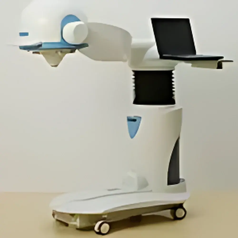
| Brand | Hamamatsu Photonics (BHP) |
|---|---|
| Model | BHP6602 |
| Type | Compact Planar Gamma Camera for Small-Animal and Human Thyroid/Parathyroid Imaging |
| Origin | Hebei, China |
| Regulatory Approvals | NMPA (ex-SFDA) Certified, FDA 510(k) Cleared |
| Field of View (FOV) | ≥150 mm |
| Energy Range | 70–364 keV (optimized for ⁹⁹ᵐTc, ¹²³I, ¹³¹I) |
| Intrinsic Spatial Resolution | ≤3.8 mm FWHM |
| Maximum Count Rate | ≥150 kcps |
| Intrinsic Uniformity | ≤±3.0% integral, ≤±0.5% differential |
| Intrinsic Spatial Linearity Error | ≤0.4 mm |
| Dimensions (L×W×H) | 1192 × 804 × 1268 mm (low position) / 1468 mm (high position) |
| Weight | 147 kg |
| PACS Integration | Yes |
| Software Interface | Full Chinese GUI with quantitative analysis modules for thyroid area/weight, renal time-activity curves, gated blood pool, and static bone imaging |
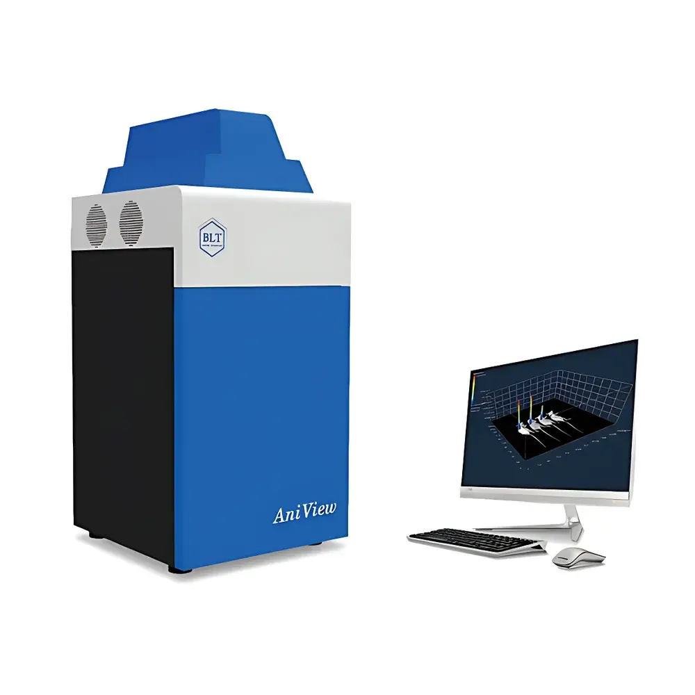
| Brand | BioLight |
|---|---|
| Model | AniView100 |
| Imaging Modality | Optical (Bioluminescence, Fluorescence, Cerenkov, Upconversion Fluorescence), Optional X-ray |
| CCD Sensor | Deep-cooled scientific-grade CCD, -100°C operating temperature |
| Filter Capacity | Up to 20 excitation LEDs + 10 emission filters |
| Software | Quantitative analysis with radiance calibration (p/s/cm²/sr), automated focus & protocol execution |
| Compliance | Designed for GLP-compliant preclinical research environments |
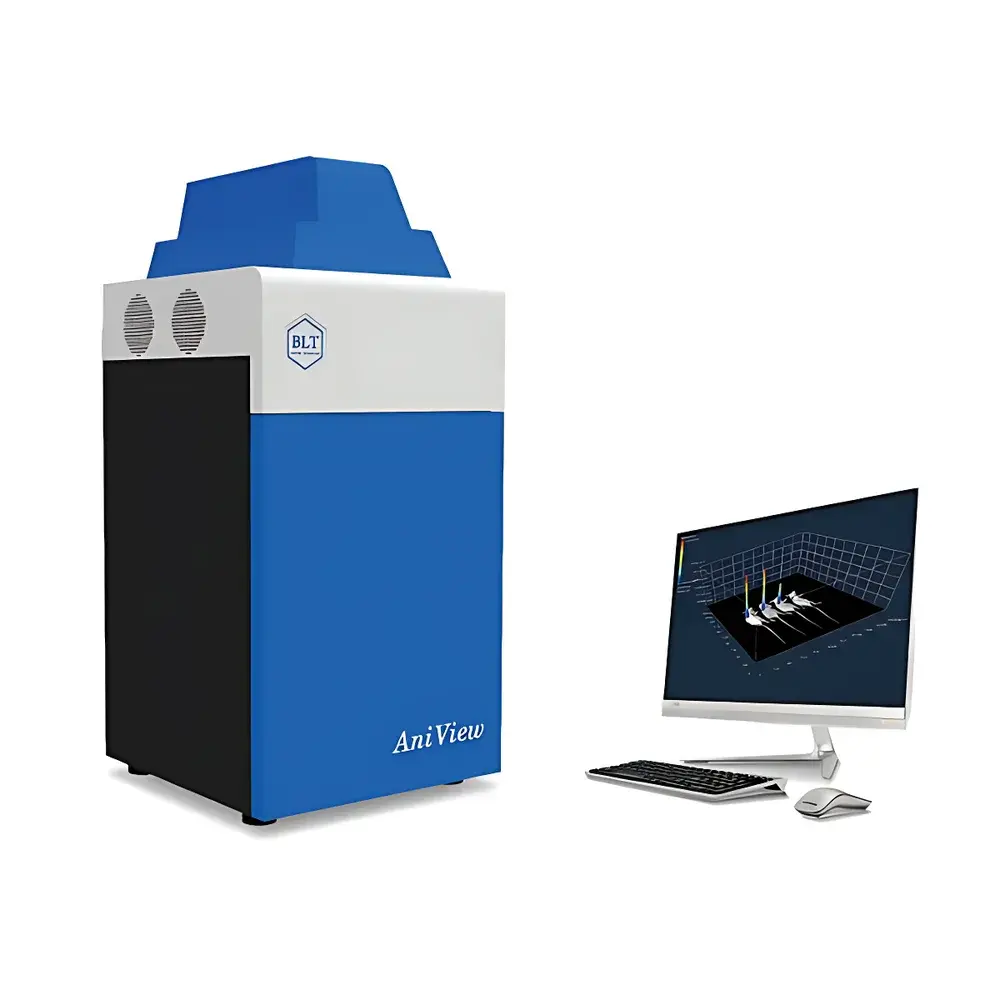
| Brand | BioLight |
|---|---|
| Origin | Guangdong, China |
| Model | PlantView100 |
| Imaging Modality | Optical Bioluminescence & Fluorescence Imaging |
| Maximum Field of View | 280 mm × 280 mm |
| Camera | Dual-cooled scientific-grade CCD (−100 °C), QE > 95% |
| Excitation Sources | Up to 20 narrowband high-power LEDs |
| Emission Filters | Up to 10 precision OD6 bandpass filters |
| Optional Modules | Side-view imaging module, X-ray imaging module, Chlorophyll fluorescence module, Light simulation module (4-channel LED: blue/white/red/NIR), Cell labeling quantification module (tube & 96-well formats) |
| Software | GLP-compliant, audit-trail enabled, auto-calibrated photon flux quantification (p/s/cm²/sr), raw + processed data segregation, export to CSV/Excel with embedded metadata |
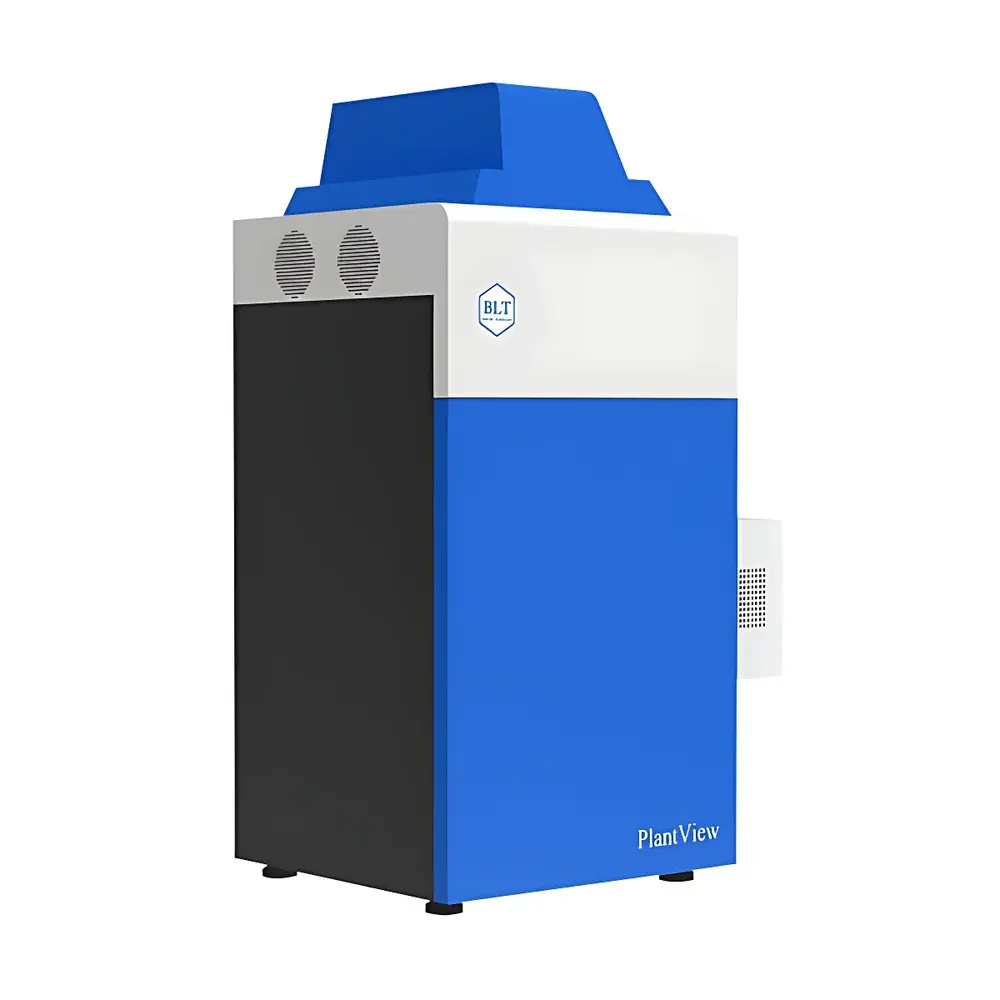
| Brand | BioLight |
|---|---|
| Origin | Guangdong, China |
| Manufacturer Type | OEM Manufacturer |
| Origin Category | Domestic |
| Model | PlantView600 |
| Price | Upon Request |
| Resolution | 2688 × 2200 |
| Pixel Size | 4.54 µm × 4.54 µm |
| Field of View | 280 mm × 230 mm |
| Brand | Biopharma |
|---|---|
| Origin | United Kingdom |
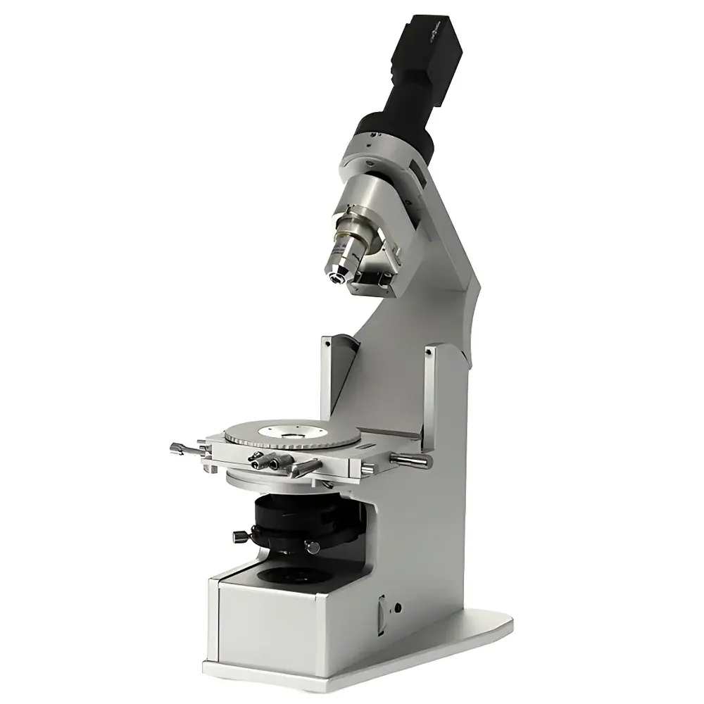
| Model | LyoStat5 |
| Application | In-situ freeze-drying process visualization and thermal transition analysis |
| Cooling System | Automated dual-pump liquid nitrogen system with 2 L Dewar and insulated flexible transfer lines |
| Vacuum Control | Motorized, digitally regulated vacuum valve with pressure display in mbar, kPa, and mTorr |
| Optical System | High-transmission, low-aberration modular optics with integrated high-brightness LED illumination (rated >60,000 h) |
| Imaging | USB 3.0 digital camera (1920 × 1080 resolution, 60 fps), real-time video capture and time-stamped image logging up to 80 GB storage |
| Software | LyoView™ v5.2 — fully programmable temperature ramp/hold profiles, synchronized pressure logging, event-triggered image capture, audit-trail enabled for GLP/GMP environments |
| Compliance | Designed to support ASTM F2721-22 (Standard Guide for Characterization of Freeze-Dried Protein Pharmaceuticals), ISO 22675:2021 (Pharmaceutical freeze-drying process development), and FDA 21 CFR Part 11-compliant data integrity workflows |
| Brand | Bioptechs |
|---|---|
| Origin | USA |
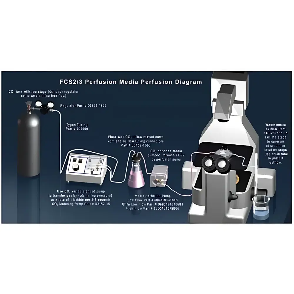
| Model | Delta T |
| Microscope Type | Upright Microscope |
| Sample Chamber | 35 mm Standard Petri Dish with Bonded 170 µm Glass Coverslip |
| Temperature Control Method | ITO (Indium Tin Oxide) Transparent Heated Layer on Coverslip Bottom |
| Temperature Feedback | External Thermistor |
| Thermal Response Rate | ~0.1 °C/s |
| Compatible Imaging Modes | Brightfield, Darkfield, Polarized Light, Phase Contrast, DIC, HMC, Fluorescence, Multiphoton, Confocal |
| Coverslip Options | Stress-Free Glass, Variable Thickness (Including High-NA Optimized) |
| Adapter Support | Brain Slices, Monolayer Cultures, Tissue Explants, Small-Area Cell Preparations |
| Compliance Context | Designed for GLP/GMP-aligned live-cell imaging workflows requiring thermal stability and environmental reproducibility |
| Brand | Biopticon |
|---|---|
| Origin | USA |
| Manufacturer Type | Authorized Distributor |
| Product Category | Imported Instrument |
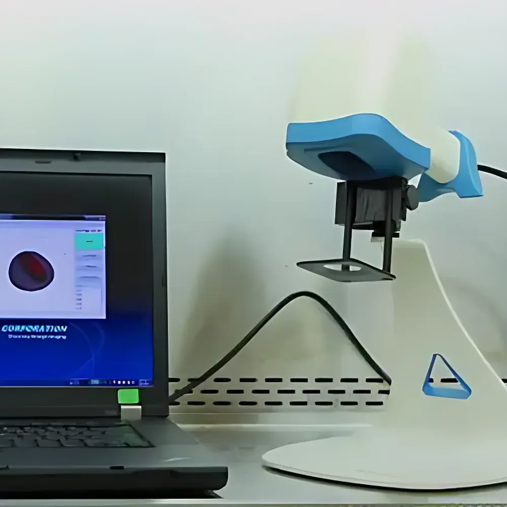
| Model | TumorImager 2 |
| Instrument Type | Optical 3D Surface Imaging System |
| Scan Resolution | 480 × 640 pixels |
| Scan Speed | <1 s per acquisition |
| Field of View | 30 mm × 20 mm |
| Sample Capacity | 1 animal per scan |
| Z-Axis Resolution | 50 µm |
| Interface | USB 3.0 |
| Output Formats | JPG (2D), OBJ & PLY (3D surface mesh) |
| Operating Modes | Handheld or Stand-Mounted |
| Tumor Length Limit | ≤30 mm |
| Tumor Height Limit | ≤20 mm |
| Automatic Segmentation Time | ~3 s |
| Data Storage | Local SQL database with user authentication and audit trail |
| Brand | BM Microscopes |
|---|---|
| Origin | Shanghai, China |
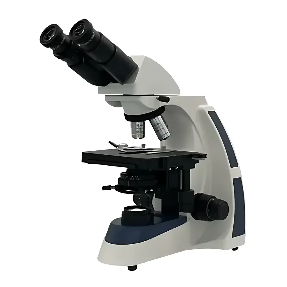
| Model | XSP-BM-17 |
| Microscope Type | Upright |
| Optical Design | Infinity-Corrected (UIS) |
| Eyepiece Configuration | Binocular |
| Standard Eyepieces | Widefield 10×/Φ22 mm |
| Optional Eyepieces | 16×/Φ15 mm |
| Objective Set | Infinity Plan Achromatic 4× (NA 0.10, WD 20.00 mm), 10× (NA 0.25, WD 13.50 mm), 40× (NA 0.65, WD 0.68 mm), 100× Oil (NA 1.25, WD 0.20 mm) |
| Total Magnification Range | 40×–1000× |
| Mechanical Stage | 140 × 160 mm, Travel Range 76 × 45 mm |
| Focusing System | Coaxial coarse/fine focus with 30 mm travel |
| Fine Focus Graduation | 2 µm |
| Condenser | Abbe condenser, NA 1.25, adjustable iris diaphragm |
| Filters | Blue and green interference filters |
| Illumination | Adjustable-intensity LED light source |
| Tube Angle | 30° inclined, 360° rotatable hinge joint |
| Compliance | Designed for ISO 10993-compatible lab environments |
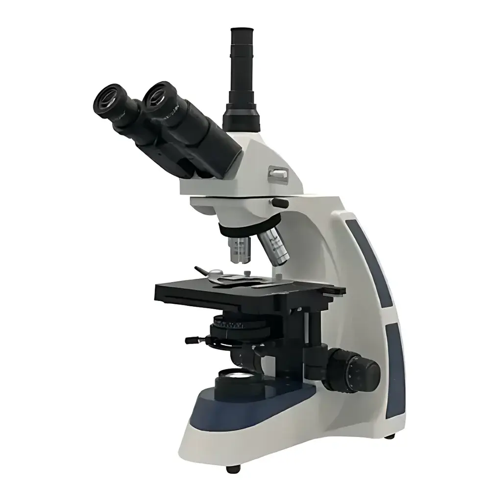
| Brand | BM Optical |
|---|---|
| Origin | Shanghai, China |
| Microscope Type | Upright |
| Configuration | Trinocular |
| Optical Design | UIS Infinity-Corrected System |
| Eyepieces | Widefield 10×/Φ22 mm (standard), optional 16×/Φ15 mm |
| Objectives | Infinity-Corrected Achromatic, 4×/0.10, 10×/0.25, 40×/0.65, 100× oil/1.25 |
| Total Magnification Range | 40×–1000× |
| Mechanical Tube Length | ∞ |
| Stage | 140 × 160 mm mechanical stage with 76 × 45 mm travel |
| Focus Mechanism | Coaxial coarse/fine focus, fine focus graduation: 2 µm |
| Condenser | Abbe condenser, NA 1.25, adjustable iris diaphragm |
| Filters | Blue and green interference filters |
| Illumination | Adjustable-intensity LED light source |
| Compliance | Designed to meet ISO 10934-1 (Microscopes — Nomenclature of components) and GB/T 2985–2019 (Biological microscopes — Requirements and test methods) |
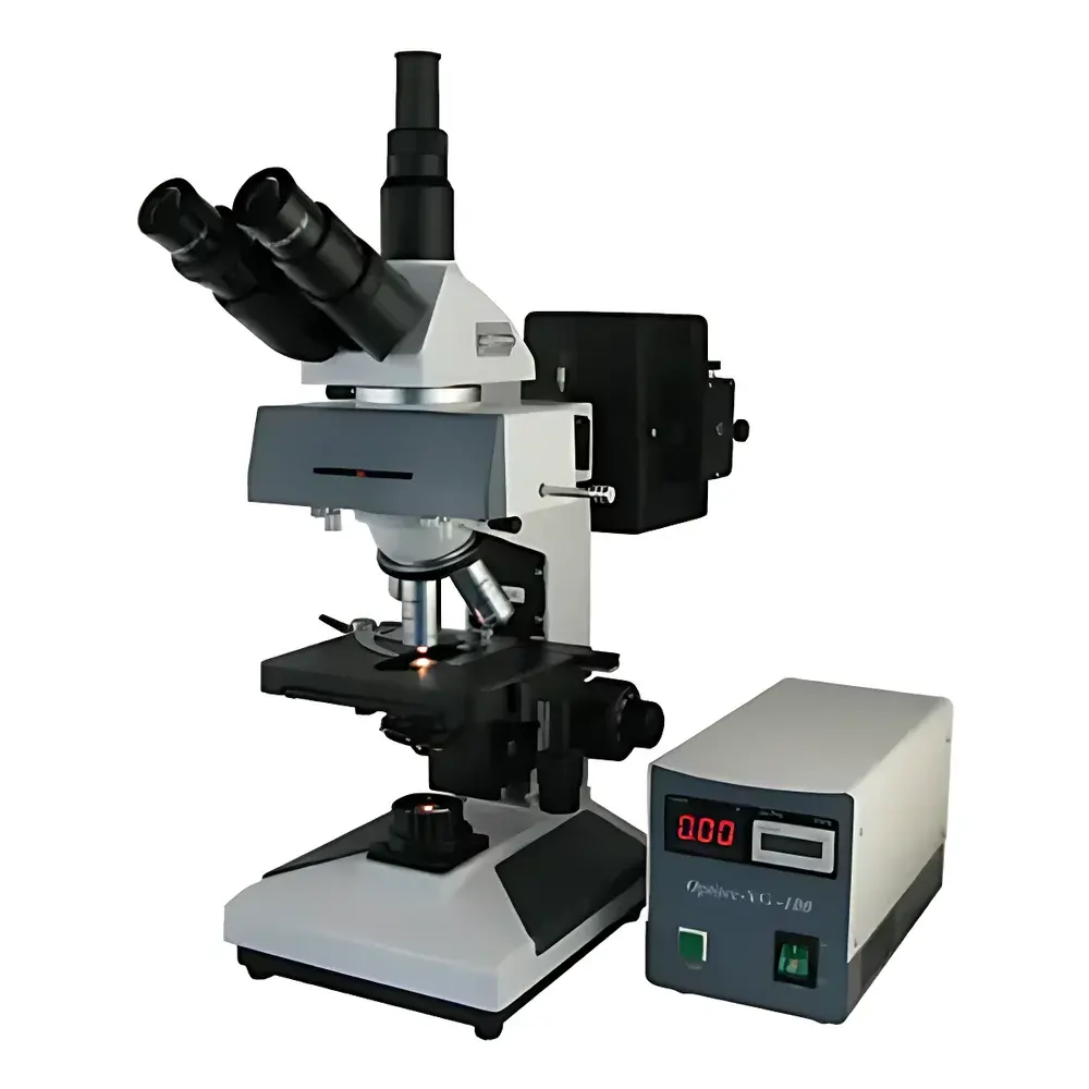
| Brand | BM |
|---|---|
| Origin | Shanghai, China |
| Model | XSP-BM-13C |
| Instrument Type | Upright Fluorescence Microscope |
| Excitation Source | Mercury Arc Lamp (100 W) with B/G Filter Sets |
| Transmitted Illumination | 6 V / 20 W Halogen Lamp |
| Eyepieces | 10× / Ø20 mm (standard), optional 16× / Ø15 mm |
| Objective System | Infinity-Corrected Fluorescence Objectives (4×, 10×, 40×, 100× oil, NA up to 1.25) |
| Mechanical Stage | 180 × 150 mm platform, 75 × 50 mm travel range |
| Condenser | Adjustable Abbe condenser, NA 1.25 with iris diaphragm |
| Observation Head | 30° inclined binocular head with 360° rotation capability |
| Focusing Mechanism | Coaxial coarse/fine focus, 30 mm vertical travel, fine focus graduation 0.002 mm, tension adjustment and limit stop |
| Fluorescence Filters | Blue (B), Green (G), Yellow (Y) excitation/emission sets |
| Interpupillary Distance | 55–75 mm |
| Tube Factor | 1× (standard infinity system) |
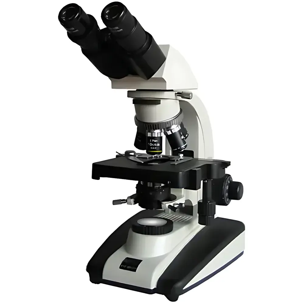
| Brand | BM |
|---|---|
| Origin | Shanghai, China |
| Microscope Type | Upright Microscope |
| Eyepiece Configuration | Binocular |
| Optical System | UIS Infinity-Corrected |
| Objective Lenses | 4×/0.10, 10×/0.25, 40×/0.65 (Spring), 100×/1.25 (Oil Immersion) |
| Eyepieces | WF10×/22 mm, WF16×/15 mm |
| Total Magnification Range | 40×–1600× |
| Mechanical Stage | Dual-layer, 155 × 145 mm, 80 × 50 mm Travel |
| Focus Mechanism | Coaxial Coarse/Fine Adjustment, 30 mm Travel, Fine Focus Graduation: 2 µm |
| Condenser | Abbe N.A. 1.25 with Centering Adjustment and Iris Diaphragm |
| Illumination | 6 V / 20 W Halogen Lamp, Köhler Illumination, Continuously Variable Brightness Control |
| Filter Set | Blue, Green, Yellow |
| Included Accessories | Immersion Oil, Spare Bulb (6 V / 20 W), Fuse (1 A), Power Cord, User Manual, Certificate of Conformance, Warranty Card |
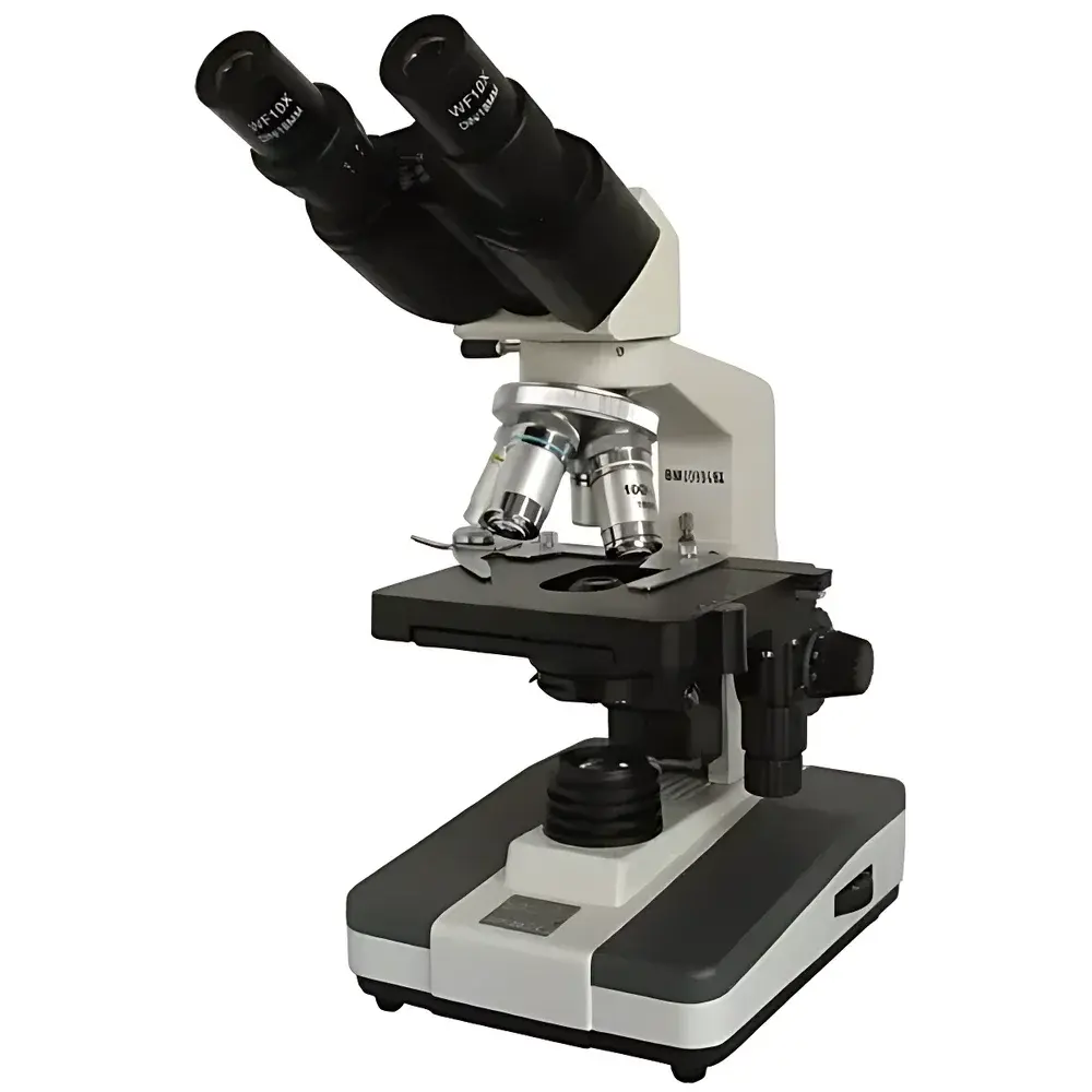
| Brand | BM |
|---|---|
| Origin | Shanghai, China |
| Microscope Type | Upright |
| Eyepiece Configuration | Binocular |
| Mechanical Tube Length | 160 mm |
| Objective Lenses | 4× (NA 0.10, WD 37.5 mm), 10× (NA 0.25, WD 7.31 mm), 40× (NA 0.65, WD 0.63 mm), 100× oil immersion (NA 1.25, WD 0.18 mm) |
| Eyepieces | Wide-field 10×/Φ18 mm and 16×/Φ11 mm |
| Total Magnification Range | 40×–1600× |
| Stage | Mechanical 130×130 mm with 70×30 mm travel and 0.1 mm vernier scale |
| Interpupillary Adjustment | 55–75 mm |
| Focusing System | Coaxial coarse/fine focus with 15 mm travel |
| fine focus graduation | 0.002 mm |
| Condenser | Abbe condenser, NA 1.25, adjustable iris diaphragm |
| Filters | Blue, green, yellow |
| Illumination | 6 V / 20 W halogen or LED lamp, continuously variable brightness |
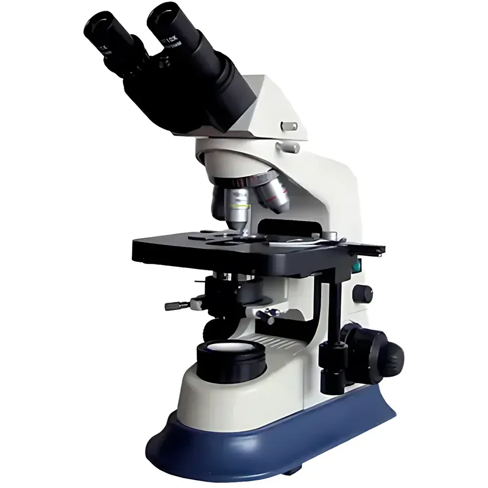
| Brand | BM |
|---|---|
| Origin | Shanghai, China |
| Microscope Type | Upright Biological Microscope |
| Optical Design | UIS Infinite Optical System |
| Eyepiece Configuration | Binocular, 30° Inclined, 360° Rotatable Head |
| Eyepieces | Widefield 10×/Φ22 mm and 16×/Φ15 mm |
| Objectives | Infinity-Corrected Achromatic Plan Objectives (4×/0.10, 10×/0.25, 40×/0.65, 100× oil/1.25) |
| Mechanical Tube Length | 160 mm |
| Total Magnification Range | 40×–1600× |
| Stage | Mechanical XY Translation Platform (175 × 150 mm, 80 × 50 mm Travel, 0.1 mm Vernier Scale) |
| Interpupillary Adjustment | 55–75 mm |
| Focus Mechanism | Coaxial Coarse/Fine Focus (30 mm Travel, Fine Focus Graduation: 2 µm) |
| Condenser | Abbe Condenser, N.A. 1.25, Centerable, with Iris Diaphragm |
| Filters | Blue, Green, Yellow |
| Illumination | 6 V / 20 W Halogen Lamp, Continuously Variable Brightness Control |
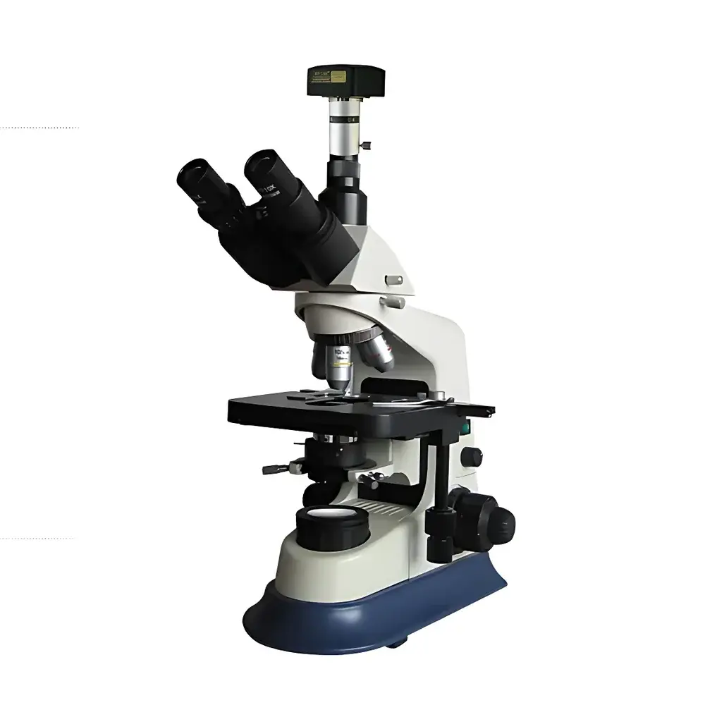
| Brand | BM |
|---|---|
| Origin | Shanghai, China |
| Model | XSP-BM-30AD |
| Microscope Type | Computer-Integrated Biological Microscope |
| Eyepiece Configuration | Binocular |
| Optical System | Infinity-Corrected UIS (Universal Intermediate System) |
| Eyepieces | Widefield Plan 10×/Φ22 mm and 16×/Φ15 mm |
| Objectives | Infinity-Corrected Achromatic Plan Objectives (4×/0.10, 10×/0.25, 40×/0.65, 100× oil/1.25) |
| Total Magnification Range | 40×–1600× |
| Mechanical Stage | 175 × 150 mm with 80 × 50 mm travel and 0.1 mm vernier scale |
| Interpupillary Adjustment | 55–75 mm |
| Focus Mechanism | Coaxial coarse/fine focusing (30 mm travel), fine focus graduation: 0.002 mm, built-in stop and tension control |
| Condenser | Abbe condenser, N.A. 1.25, centerable and iris diaphragm adjustable |
| Filters | Blue, green, yellow interference filters |
| Illumination | 6 V / 20 W halogen lamp with Köhler illumination and continuous brightness control |
| Digital Imaging System | 5 MP CMOS camera with MCL-Z C-mount adapter |
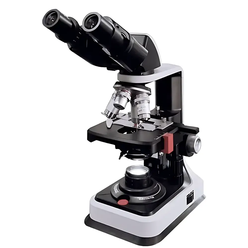
| Brand | BM |
|---|---|
| Origin | Shanghai, China |
| Microscope Type | Upright Biological Microscope |
| Eyepiece Configuration | Binocular |
| Mechanical Tube Length | 160 mm |
| Objective Lenses | 4× (NA 0.10, WD 37.5 mm), 10× (NA 0.25, WD 7.31 mm), 40× (NA 0.65, WD 0.63 mm), 100× Oil (NA 1.25, WD 0.18 mm) |
| Eyepieces | Widefield 10×/Φ18 mm (standard), optional 16×/Φ11 mm |
| Total Magnification Range | 40×–1600× |
| Stage | Dual-layer mechanical stage (110 × 120 mm, travel 70 × 30 mm) |
| Interpupillary Adjustment | 50–75 mm |
| Focus Mechanism | Coaxial coarse/fine focusing, fine focus graduation 0.02 mm |
| Condenser | Abbe condenser, NA 1.25, adjustable iris diaphragm |
| Filter | Blue filter included |
| Illumination | Adjustable-intensity LED light source with integrated Type-C rechargeable power module and onboard cable management |
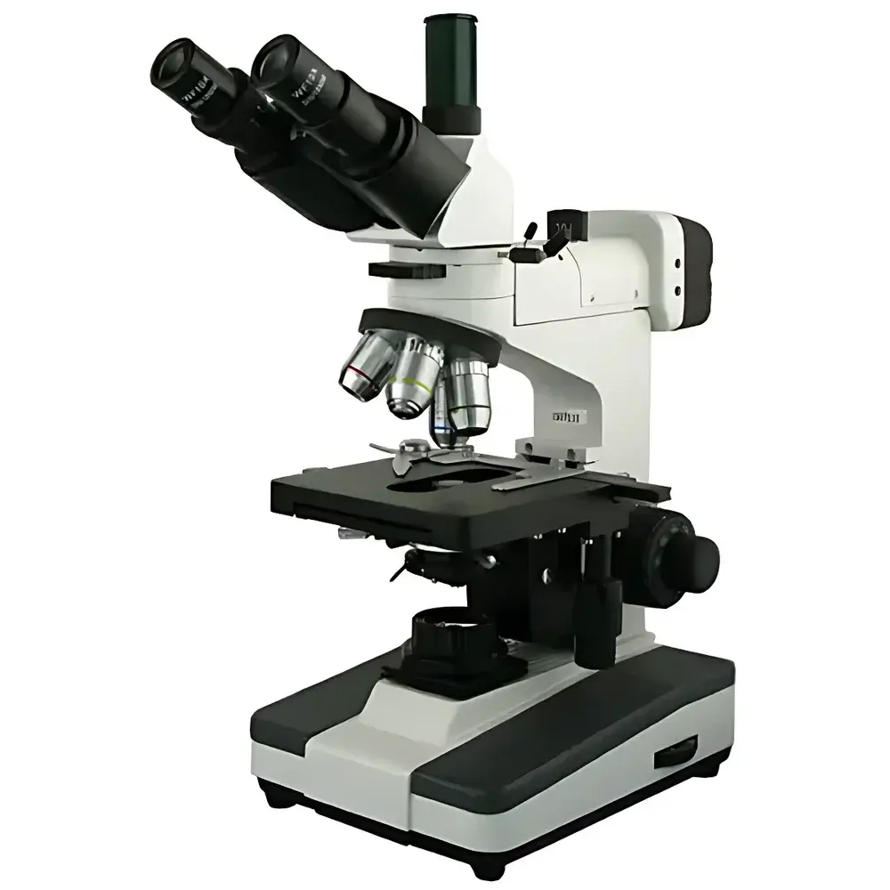
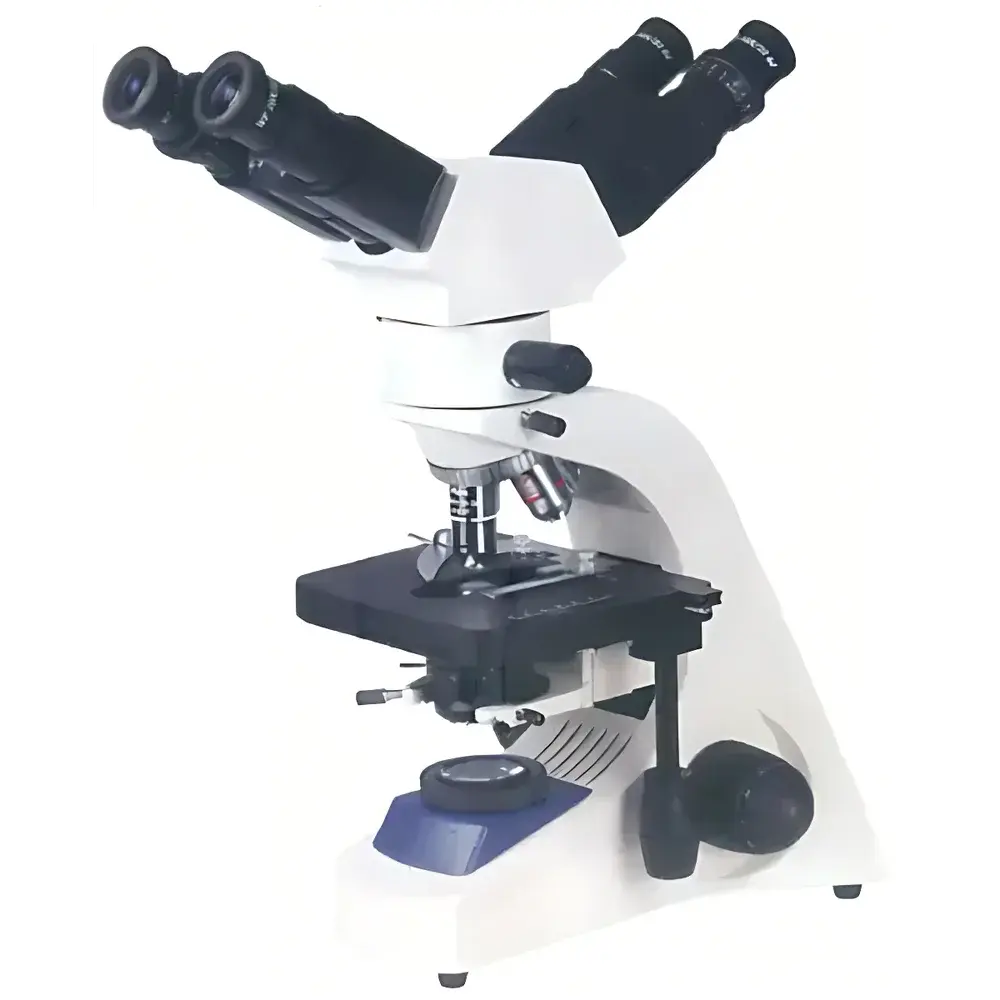
| Brand | BM |
|---|---|
| Origin | Shanghai, China |
| Model | BM-12 |
| Type | Trinocular Digital Biological Microscope |
| Illumination | Dual-mode (Transmitted & Reflected Halogen, 6V/20W) |
| Eyepiece Port | Trinocular |
| Objective Mount | Standard DIN 4X–100X Oil |
| Condenser | NA 1.25 Abbe with Iris Diaphragm & Centering Mechanism |
| Focusing | Coaxial Coarse/Fine Adjustment (Fine Graduation: 2 µm) |
| Stage | Dual-layer Mechanical Stage (140 × 140 mm, 75 × 40 mm Travel) |
| Polarizer System | Rotatable Analyzer (0–360°), Removable Polarizer & Analyzer |
| Filters | Insert-type Green/Blue/Yellow |
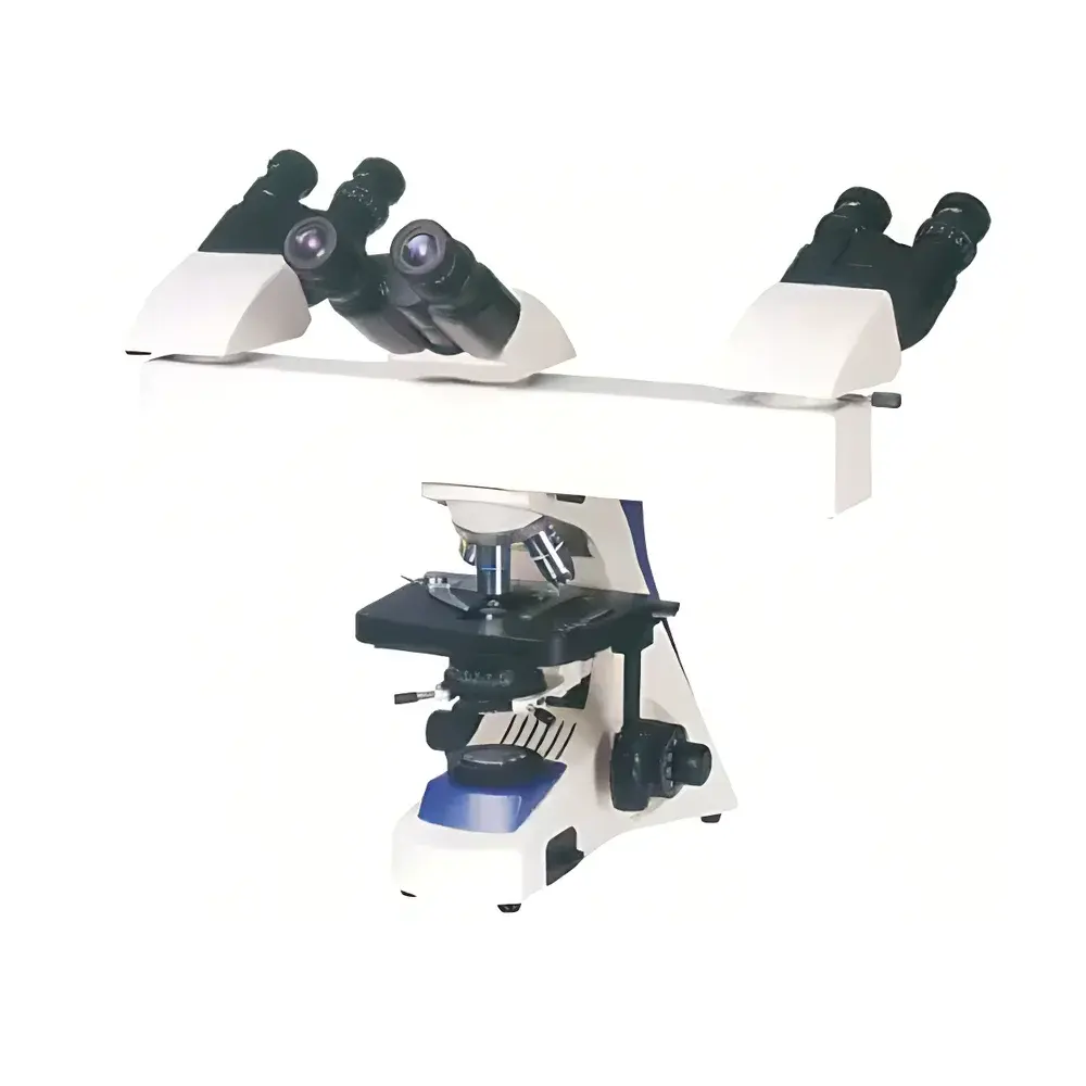
| Brand | BM |
|---|---|
| Origin | Shanghai, China |
| Model | BM-18F2 |
| Microscope Type | Inverted Teaching Microscope |
| Eyepiece Configuration | Binocular |
| Optical System | Infinity-Corrected |
| Illumination | 6V/20W Halogen with Köhler Illumination |
| Objective Lenses | 4×, 10×, 40×(S), 100×(S) Oil Immersion (Achromatic Infinity-Corrected) |
| Eyepieces | Widefield 10×/22 mm |
| Stage | Double-layer Mechanical Stage (180 × 150 mm |
| Condenser | NA 1.25 Abbe Condenser with Iris Diaphragm and Filter Holder |
| Focus Mechanism | Coaxial Coarse/Fine Focus (Fine Focus Graduation: 2 µm) |
| Interpupillary Adjustment Range | 55–75 mm |
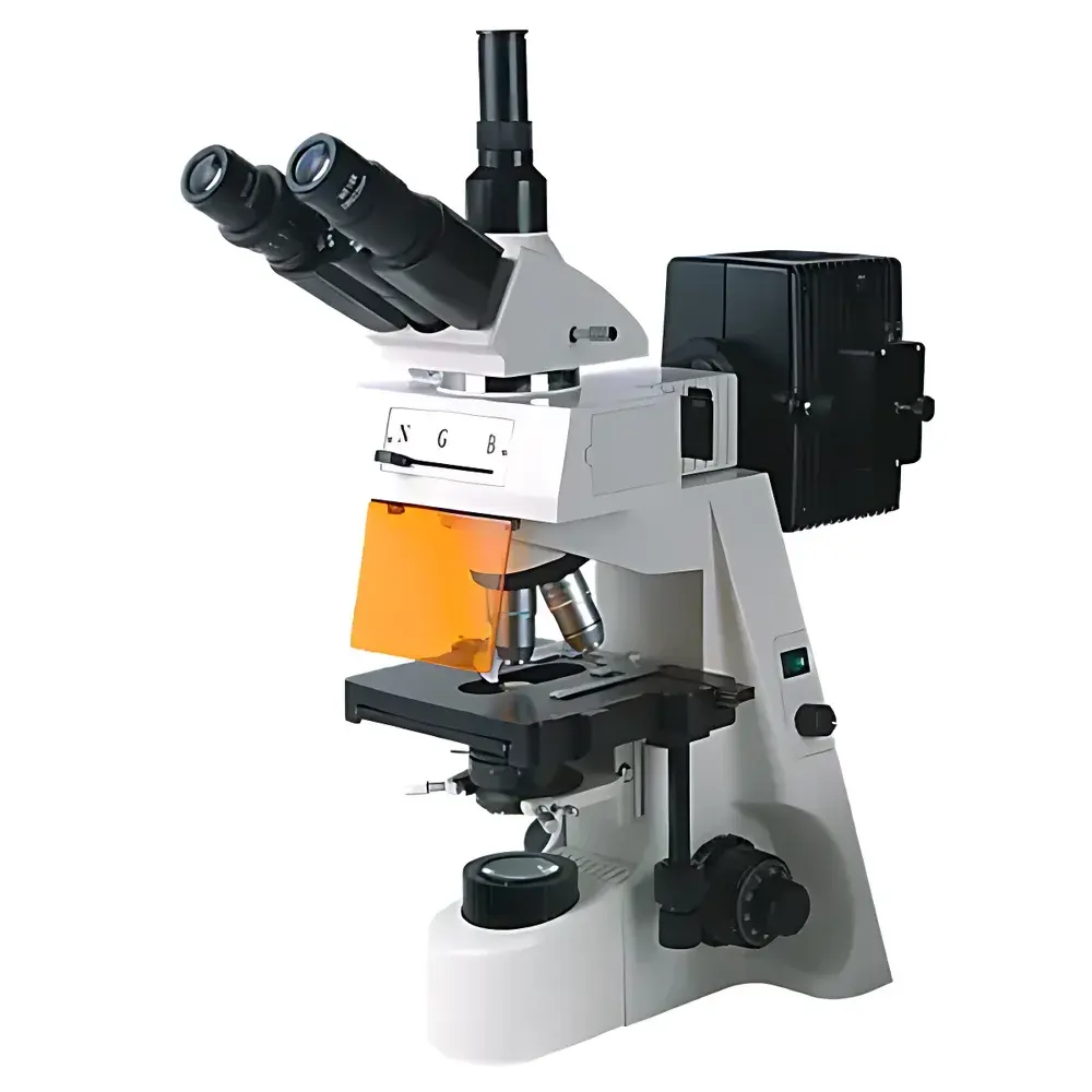
| Brand | BM |
|---|---|
| Origin | Shanghai, China |
| Model | BM-18F3 |
| Microscope Type | Upright Biological Microscope |
| Observation Head | Hinged Trinocular Head with Interpupillary Adjustment (55–75 mm) |
| Eyepieces | Widefield 10×/22 mm |
| Objectives | Infinity-Corrected Achromatic Objectives — 4×, 10×, 40×(S), 100×(S) Oil Immersion |
| Stage | Dual-layer Mechanical Stage (180 × 150 mm) |
| Travel Range | 75 × 50 mm |
| Condenser | Abbe Condenser, NA 1.25, with Iris Diaphragm and Filter Holder |
| Focus Mechanism | Coaxial Coarse/Fine Focus |
| Fine Focus Graduation | 2 µm |
| Illumination | 6 V / 20 W Halogen Lamp (110 V / 220 V Compatible) |
| LED Pointer System | Adjustable Brightness, Arrow-Shaped Cursor, Fully Movable Within Field of View |
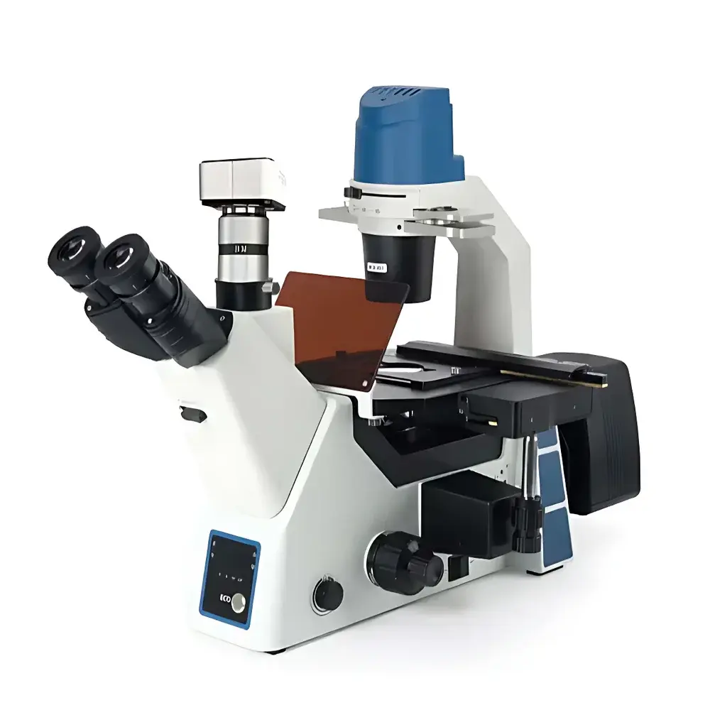
| Brand | BM |
|---|---|
| Origin | Shanghai, China |
| Model | BM-19AY |
| Instrument Type | Upright Fluorescence Microscope |
| Excitation Source | 100 W High-Pressure Mercury Lamp |
| Observation Head | Trinocular, 30° Inclined, 360° Rotatable |
| Eyepieces | Wide-Field Plan Eyepieces (10×/Φ22 mm) |
| Objectives | Infinity-Corrected Fluorescence Plan Achromatic Objectives (4×, 10×, 40×, 100× oil) |
| Illumination | Epi-Fluorescence (U, V, B, G filter sets) + Transmitted Köhler Illumination (12 V/30 W Halogen) |
| Focusing | Coaxial Coarse/Fine Manual Focus (30 mm Travel, 0.002 mm Fine Graduation) |
| Stage | 180 × 150 mm Mechanical Stage with 75 × 50 mm Travel and 0.1 mm Vernier Scale |
| Tube Lens Focal Length | 200 mm |
| Optical System | Infinity-Corrected (∞) |
| Regulatory Status | Not a Medical Device |
| Brand | BM-Beam |
|---|---|
| Origin | Shanghai, China |
| Model | BM-40YD |
| Instrument Type | Inverted Fluorescence Microscope |
| Research Grade Classification | Yes |
| Fluorescence Filter Cubes | UV1 (385 nm center), B1 (470 nm center), G1 (560 nm center), R1 (630 nm center, optional) |
| Light Source | LED-based, 385 nm / 470 nm / 560 nm triple-channel excitation |
| Control Interface | PC software-controlled fluorescence filter turret and illumination |
| Focusing Mechanism | Coaxial coarse/fine manual focus with 0.002 mm fine adjustment resolution |
| Condenser | NA 0.3, long working distance (WD = 72 mm), removable, phase-adjustable slider with dual centering knobs |
| Observation Head | 45° inclined hinge-type trinocular, 360° rotatable, interpupillary distance 50–75 mm, dual-mode beam splitter (100% eyepiece / 100% camera) |
| Stage | Three-layer mechanical stage, 215 × 250 mm platform, 120 × 80 mm travel range |
| Objective Lenses | Infinity-corrected semi-apochromatic plan fluor objectives (4×/0.13, WD 18.5 mm |
| Optical System | Infinity-corrected semi-plan achromat optical design |
| Illumination | Adjustable warm/cool white transmitted LED, independent intensity control per channel, IR motion sensing, LED fluorescence status indicator |
| Fluorescence Light Engine | Four-position motorized filter turret with servo-driven gear transmission, integrated LED driver for simultaneous or sequential excitation control |
| Brand | Bruker |
|---|---|
| Origin | USA |
| Manufacturer Type | Authorized Distributor |
| Origin Category | Imported |
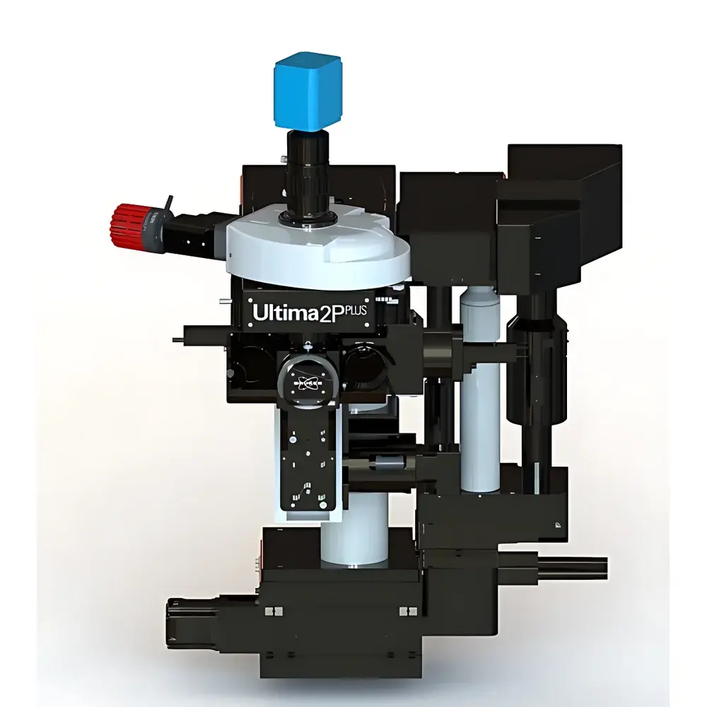
| Model | 2PPlus |
| Price Range | USD $380,000–$640,000 (est.) |
| Instrument Type | Optical In Vivo Imaging System |
| Scanning Resolution | 2048 × 2048 pixels |
| Maximum Frame Rate | 1300 fps |
| Field of View | 1.9 mm × 1.9 mm (with 16× objective) |
| Sample Capacity | 1 specimen per acquisition session |
| Excitation Wavelength Range | 400–1700 nm |
| Multiphoton Capability | Two-photon and three-photon imaging supported |
| Scan Modes | Galvo-based raster, spiral, and resonant scanning |
| Objective Compatibility | Motorized and manual multi-axis rotating objectives (single- and multi-axis) |
| Optional Modules | Full-field LED illumination, 1P/2P uncaging, spatial light modulator (SLM)-based patterned excitation |
| Brand | Bruker |
|---|---|
| Origin | USA |
| Manufacturer Type | Authorized Distributor |
| Origin Category | Imported |
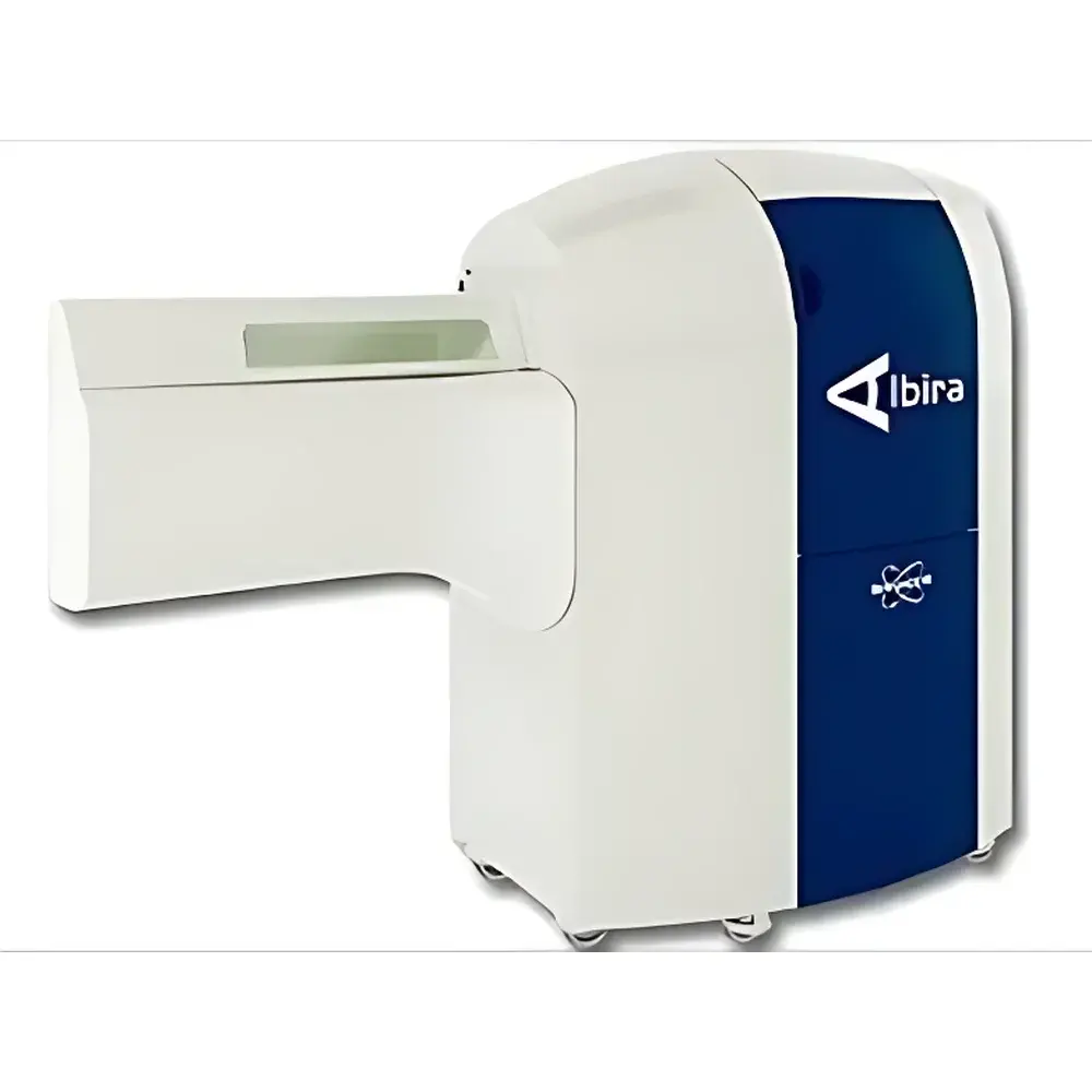
| Model | Albira Si |
| Price Range | USD 1,100,000 – 1,380,000 (based on FOB US pricing and typical import duty/tax structure) |
| Instrument Type | Trimodal Molecular Imaging System |
| Brand | Bruker |
|---|---|
| Origin | Germany |
| Manufacturer Type | Authorized Distributor |
| Origin Category | Imported |
| Model | BioSpec |
| Instrument Type | Nuclear Magnetic Resonance (NMR) Imaging System |
| Field Strength Options | 4.7 T, 7.0 T, 9.4 T, 11.7 T, and 15.2 T configurations available |
| Cryogenic Probe Technology | MRI CryoProbe® |
| Magnet Type | Ultra-Shielded Resistive (USR) or Superconducting Magnets |
| Regulatory Compliance | CE-marked, ISO 13485-certified manufacturing processes, compliant with IEC 62304 for medical device software lifecycle management |
| Brand | Bruker |
|---|---|
| Origin | Germany |
| Manufacturer Type | Authorized Distributor |
| Origin Category | Imported |
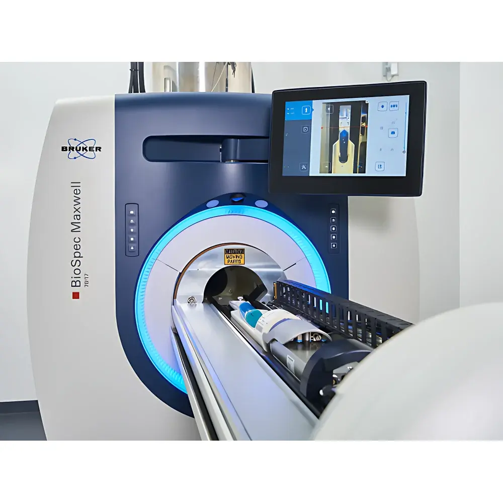
| Model | Maxwell |
| Instrument Type | Preclinical Magnetic Resonance Imaging (MRI) System |
| Energy Resolution | 1000 (arbitrary units, typical for spectral stability metric) |
| Spatial Resolution | 0.05 mm (isotropic, in vivo) |
| Scan Time per Sequence | <1 min (typical for rapid anatomical protocols) |
| Simultaneous Sample Capacity | 2 animals |
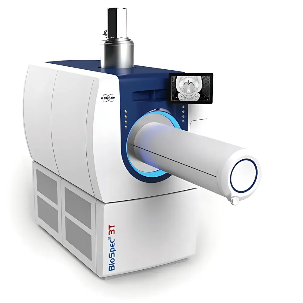
| Brand | Bruker |
|---|---|
| Origin | Germany |
| Model | BioSpec® 3T |
| Field Strength | 3 Tesla |
| Instrument Type | Preclinical Magnetic Resonance Imaging (MRI) and Magnetic Resonance Spectroscopy (MRS) System |
| Bore Diameter | 180 mm |
| RF Coil Options | >30 dedicated coils for mouse/rat head, brain, heart, body, multinuclear, and functional applications |
| Gradient Strength | Up to 900 mT/m |
| Gradient Linearity | ±3.5% within 50 mm DSV |
| Shim System | 6 high-order shim coils + high-power shim power supplies (up to 3750 Hz/cm² in XZ/YZ) |
| Magnetic Field Homogeneity | ±0.1 ppm within 50 mm DSV |
| Magnet Hold Time | >4 hours after power loss or coolant interruption |
| Software Platform | ParaVision® with >1000 optimized pulse sequences including IntraGate™, UTE, ZTE, and non-ECG-triggered cardiac imaging |
| Multi-Modal Compatibility | PET, optical imaging, CT |
| Regulatory Compliance | Designed for GLP-compliant preclinical research environments |
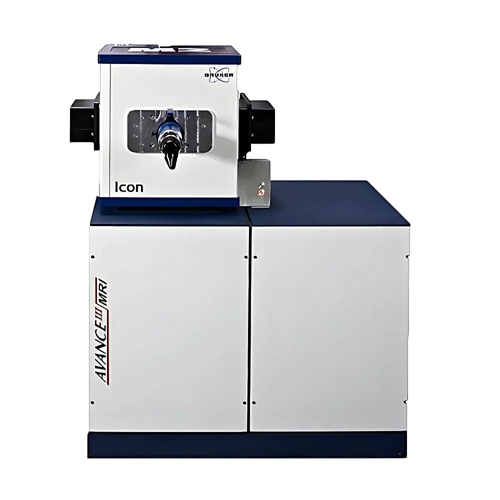
| Brand | Bruker |
|---|---|
| Origin | Germany |
| Manufacturer Type | Authorized Distributor |
| Origin Category | Imported |
| Model | ICON |
| Instrument Type | Magnetic Resonance Imaging (MRI) System |
| Field Strength | 1 Tesla |
| Target Subjects | Rodents (Mice & Rats) |
| Magnet Type | Cryogen-Free Permanent Magnet (Aspect Magnet Technologies) |
| Core Platform | AVANCE III Spectrometer Architecture |
| Software Suite | ParaVision® 360 |
| Application Domain | Preclinical Molecular Imaging, Longitudinal In Vivo Studies, Educational MRI Training |
| Operating Cost Profile | Low (No Liquid Helium or Cryogen Refills Required) |
| Footprint | Benchtop (≤ 1.2 m × 0.8 m × 1.4 m) |
| Regulatory Compliance | CE Marked, ISO 13485–Compliant Design, FDA 510(k)-Cleared for Preclinical Use (K220927) |
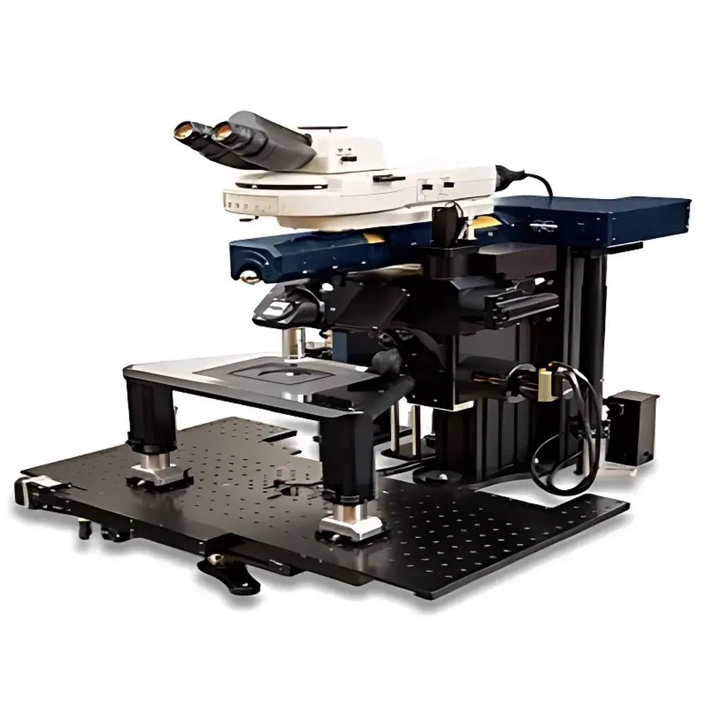
| Brand | Bruker |
|---|---|
| Origin | USA |
| Manufacturer Type | Authorized Distributor |
| Origin Category | Imported |
| Model | Investigator |
| Price Range | USD 420,000 – 700,000 |
| Instrument Type | Optical Imaging System |
| Scanning Resolution | ~700 nm |
| Maximum Frame Rate | 1300 fps |
| Field of View | 0.85 × 0.85 mm (with 16× objective) |
| Sample Capacity | 1 animal per session |
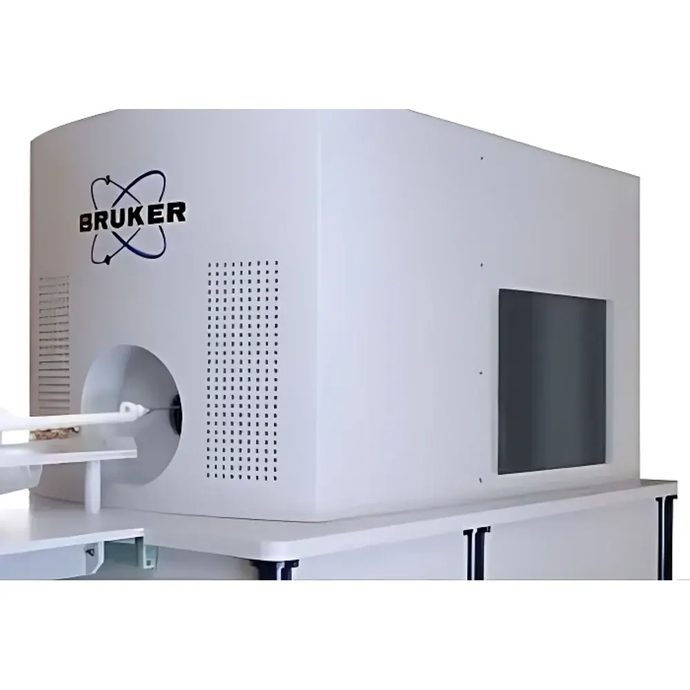
| Brand | Bruker |
|---|---|
| Origin | Germany |
| Manufacturer Type | Authorized Distributor |
| Origin Category | Imported |
| Model | MPI |
| Price Range | USD 420,000 – 700,000 |
| Instrument Type | Nuclear Imaging |
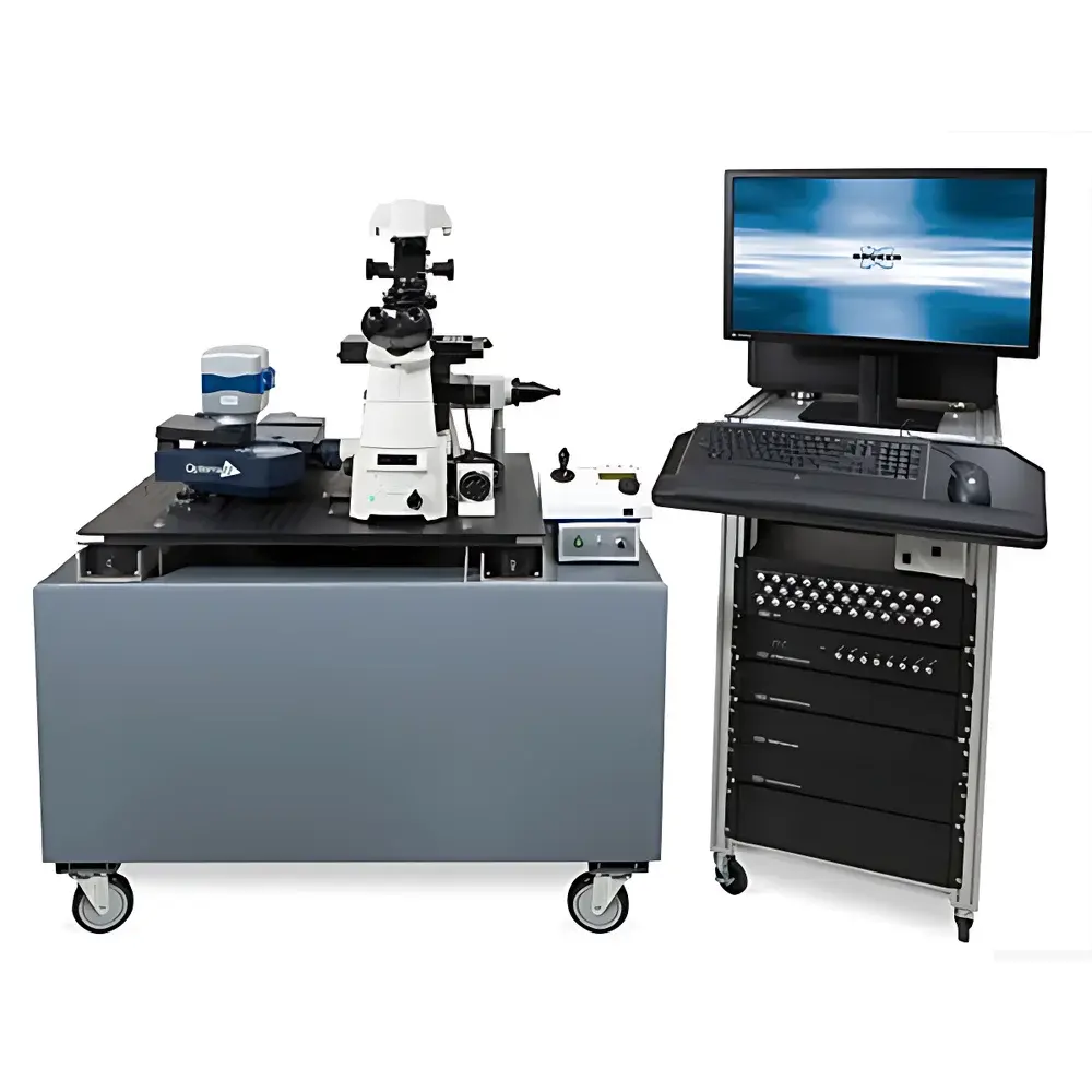
| Brand | Bruker |
|---|---|
| Origin | USA |
| Manufacturer Type | Authorized Distributor |
| Origin Category | Imported |
| Model | Opterra II |
| Price Range | USD 420,000 – 700,000 |
| Instrument Type | Optical Imaging System |
| Maximum Scan Resolution | 2048 × 2048 pixels |
| Frame Rate | up to 100 fps |
| Field of View | Objectives-dependent (e.g., 0.25 mm² at 60×, 1.0 mm² at 10×) |
| Sample Capacity | Single specimen stage with motorized XYZ + focus control |
| Detection Technology | EMCCD and sCMOS dual-path detection |
| Scanning Method | Parallel field-scanning via programmable slit/multi-spot illumination |