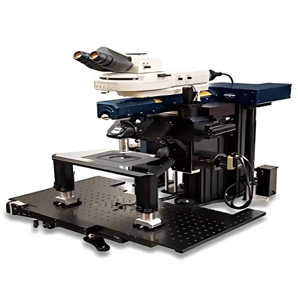
Bruker Investigator High-Performance Two-Photon In Vivo Imaging System
| Brand | Bruker |
|---|---|
| Origin | USA |
| Manufacturer Type | Authorized Distributor |
| Origin Category | Imported |
| Model | Investigator |
| Price Range | USD 420,000 – 700,000 |
| Instrument Type | Optical Imaging System |
| Scanning Resolution | ~700 nm |
| Maximum Frame Rate | 1300 fps |
| Field of View | 0.85 × 0.85 mm (with 16× objective) |
| Sample Capacity | 1 animal per session |
Overview
The Bruker Investigator is a high-fidelity two-photon in vivo imaging system engineered for deep-tissue, high-resolution structural and functional visualization in live small animals. Leveraging near-infrared (NIR) and infrared excitation lasers (typically 680–1300 nm), the system achieves nonlinear excitation of endogenous or exogenous fluorophores with minimal photodamage and scattering—enabling optical sectioning at depths exceeding 1 mm in intact brain tissue, spinal cord, lymphoid organs, and other highly scattering biological specimens. Its core optical architecture adheres to rigorous principles of multiphoton excitation microscopy (MPM), where simultaneous absorption of two low-energy photons induces fluorescence emission equivalent to that of single-photon UV/visible excitation—but with superior depth penetration and spatial confinement of excitation volume. Designed specifically for longitudinal physiological studies, the Investigator supports stable, long-duration imaging under controlled environmental conditions (temperature, humidity, gas anesthesia), preserving native tissue architecture and dynamic cellular behavior.
Key Features
- Three configurable scanning modalities: galvanometric mirror scanning for high-fidelity raster acquisition, spiral scanning for optimized signal-to-noise ratio in low-light conditions, and resonant scanning for ultra-high-speed volumetric imaging up to 1300 frames per second.
- Modular rotating objective turret supporting single-axis manual, multi-axis manual, and motorized rotation—enabling oblique-angle imaging without mechanical displacement of the specimen, critical for maintaining physiological integrity during multi-view acquisition.
- Integrated motorized microscope stage and optional mobile imaging platform, permitting precise repositioning of the optical head relative to stationary subjects—eliminating sample motion artifacts and enabling natural posture retention during chronic or awake-behavioral experiments.
- Optimized optical path design with high-transmission dichroics, low-autofluorescence objectives (e.g., 16×, 25× water-immersion), and photon-efficient detection using hybrid PMTs or GaAsP detectors calibrated for NIR spectral response.
- Robust mechanical architecture compliant with ISO 14644-1 Class 5 cleanroom-compatible installation standards; vibration-isolated optical table integration supported via standardized mounting interfaces.
Sample Compatibility & Compliance
The Investigator accommodates standard rodent models (mouse, rat) in stereotaxic or custom restraint fixtures, as well as explanted tissues (e.g., acute brain slices, tumor explants, intestinal organoids) mounted on heated perfusion chambers. It is compatible with common fluorescent indicators (GCaMP, jRGECO, tdTomato), genetically encoded sensors, and nanoparticle-based contrast agents. The system meets essential regulatory expectations for preclinical instrumentation: full alignment with GLP-compliant documentation workflows, audit-trail-enabled acquisition metadata logging (timestamp, laser power, detector gain, scan parameters), and compatibility with FDA 21 CFR Part 11–compliant electronic signature modules when integrated with validated LIMS or ELN platforms. All optical components comply with IEC 60825-1:2014 laser safety standards and are CE-marked for research use only (RUO).
Software & Data Management
Acquisition and analysis are managed through Bruker’s proprietary PrairieView software suite—built on a deterministic real-time kernel for sub-millisecond timing precision across hardware subsystems (laser modulation, scanner synchronization, analog input/output). The software supports automated Z-stack acquisition, time-lapse series with programmable interval triggers, region-of-interest (ROI) based photostimulation coupling, and offline motion correction algorithms. Raw data is saved in vendor-neutral HDF5 format with embedded metadata (MIAME/MINSEQ-compliant headers), ensuring interoperability with open-source tools such as ImageJ/Fiji, Python-based Napari, and MATLAB-based analysis pipelines. Export options include TIFF stacks, NRRD volumes, and CSV-formatted quantitative metrics (intensity, FRET ratio, calcium transient kinetics) suitable for statistical modeling in R or Python.
Applications
- Longitudinal neurovascular coupling studies in awake, head-fixed mice using cranial window preparations.
- Intravital imaging of immune cell trafficking in lymph nodes, bone marrow, or solid tumors under physiological flow conditions.
- Functional mapping of dendritic spine dynamics, synaptic plasticity, and microglial surveillance in disease models (Alzheimer’s, epilepsy, stroke).
- High-speed calcium imaging across cortical layers during sensory stimulation or behavioral tasks.
- Multi-angle structural reconstruction of vascular networks in transgenic reporter lines (e.g., Thy1-YFP, Cdh5-GFP).
FAQ
What laser wavelengths are supported by the Investigator system?
The system is optimized for tunable femtosecond Ti:Sapphire lasers (680–1080 nm) and optionally integrated OPOs extending into the mid-IR range (up to 1300 nm), enabling excitation of diverse fluorophores including IR-Dye derivatives and upconversion nanoparticles.
Can the system be upgraded for three-photon imaging?
Yes—through optional optical path modifications, including dispersion compensation modules and high-NA objectives with extended transmission beyond 1300 nm, the platform supports experimental three-photon configurations for enhanced penetration in dense tissues such as white matter tracts.
Is remote operation supported for multi-user core facilities?
Absolutely—the PrairieView software includes role-based access control, secure SSH tunneling, and VNC-compatible remote desktop protocols, allowing centralized instrument management across institutional networks while maintaining data sovereignty and HIPAA/FERPA-aligned security policies.
Does Bruker provide application support for method development?
Yes—Bruker’s Application Scientists offer on-site and virtual protocol optimization services, including optical clearing validation, anesthesia regimen calibration, and quantitative image analysis pipeline development aligned with journal-specific reporting guidelines (e.g., Nature Methods, eLife).


