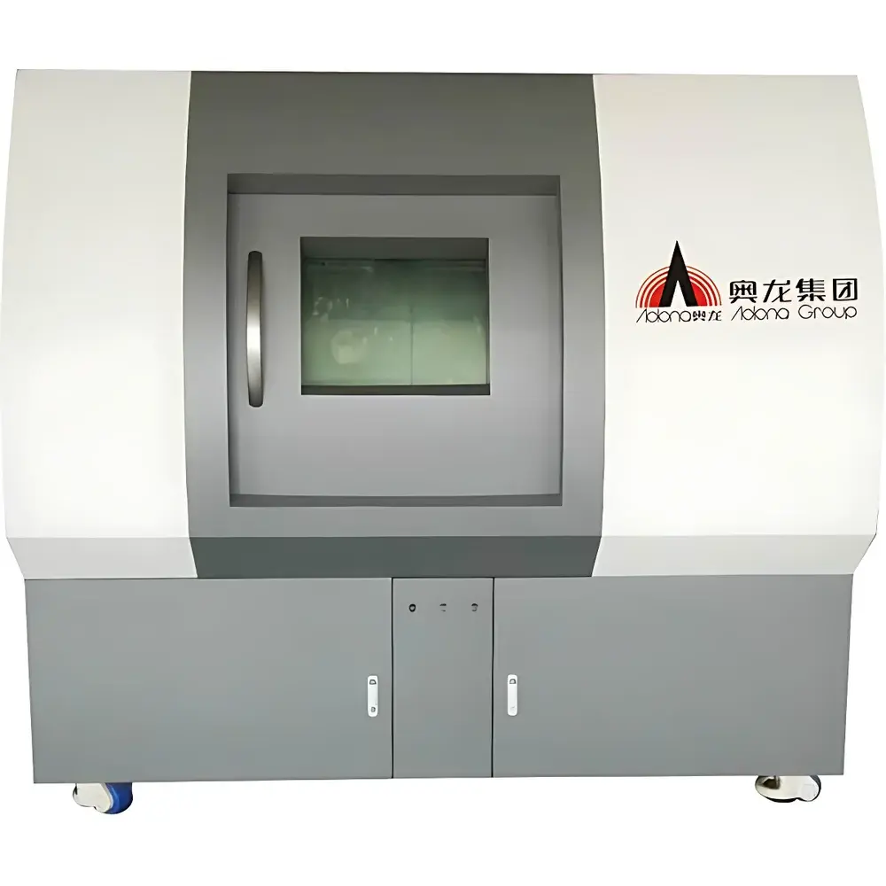
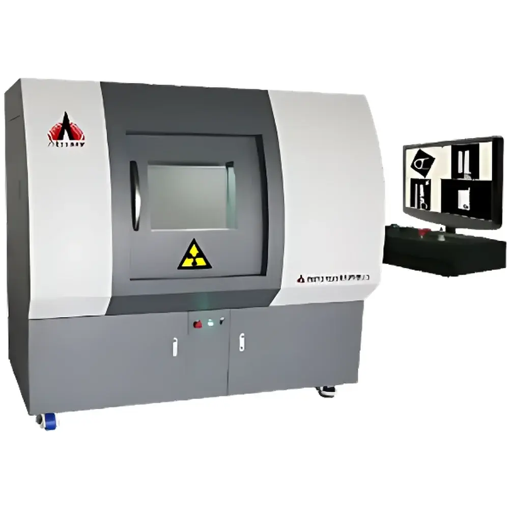
Aolong Microfocus X-ray Industrial CT System
| Brand | Aolong |
|---|---|
| Origin | Liaoning, China |
| Manufacturer Type | Manufacturer |
| Regional Classification | Domestic (China) |
| Model | Microfocus X-ray Industrial CT |
| Price Range | USD 140,000 – 280,000 (FOB) |
| Configuration | Fixed or Mobile |
| Beam Geometry | Directional |
| Minimum Focal Spot Size | 2 µm |
| Radiation Cone Angle | 30° |
| Maximum Penetration | 40 mm Al-equivalent |
Overview
The Aolong Microfocus X-ray Industrial CT System is a high-resolution, non-destructive 3D volumetric imaging platform engineered for precision internal structure analysis across demanding industrial and research environments. Based on cone-beam computed tomography (CBCT) principles, the system utilizes a microfocus X-ray source to generate high-contrast, sub-micron spatial resolution projections—enabling true 3D reconstruction of internal geometry, density distribution, and defect morphology without sample sectioning. Its core architecture integrates a rigid granite base, high-precision linear motion stages, and air-bearing rotation systems to minimize mechanical drift and ensure geometric fidelity during long-duration scans. Designed for compliance with international radiation safety standards, the system maintains an ambient dose rate below 1 µSv/h at 1 m from the enclosure—meeting IEC 61331-1 and national regulatory requirements for Class II radiation equipment.
Key Features
- Microfocus X-ray source with selectable configurations: standard 160 kV sealed-tube or optional high-energy open-tube up to 300 kV, supporting broad material penetration ranges.
- Minimum focal spot size of 2 µm—enabling high magnification imaging with theoretical spatial resolution down to 0.5 µm (JIMA-certified under optimal geometric conditions).
- Cone-beam acquisition geometry with optional helical scanning mode for extended field-of-view and improved axial resolution in elongated samples.
- Granite structural base combined with high-stiffness linear guides and air-bearing rotary stage—ensuring mechanical stability and sub-micron repeatability over thousands of scan cycles.
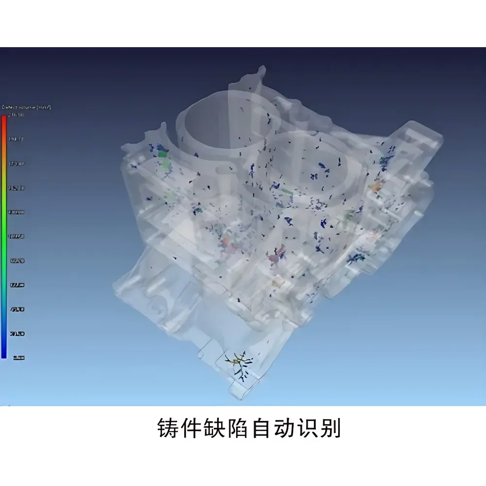
- Integrated real-time defect visualization and quantitative porosity analysis engine: automatic segmentation, color-coded volumetric rendering by defect size, and ROI-based statistical reporting.
- Low operational and maintenance cost profile: open-tube source design extends service life; modular subsystems simplify calibration and field servicing.
Sample Compatibility & Compliance
The system accommodates diverse sample geometries—from small electronic components (e.g., solder joints, MEMS packages) to castings up to Ø300 mm × H400 mm. It delivers reliable contrast for homogeneous metals (Al, Mg, Ti alloys), ceramics, polymers, geological cores, and composite laminates. Applications align with ASTM E1441 (Standard Guide for Computed Tomography), ISO 15732 (Industrial CT terminology and metrology), and support GLP/GMP traceability when paired with audit-log-enabled software modules. Radiation shielding meets GBZ 130–2020 (Chinese National Standard for X-ray Equipment Protection) and is compatible with facility-level licensing under local regulatory frameworks.
Software & Data Management
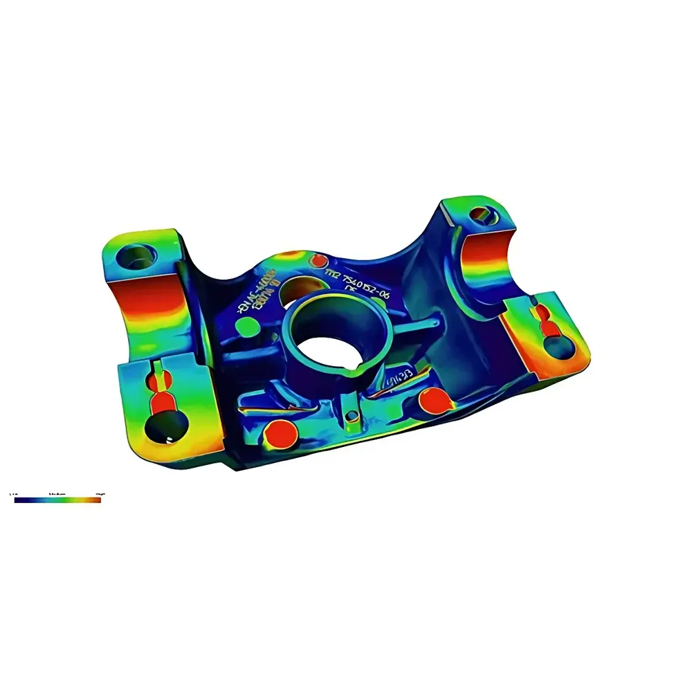
The proprietary reconstruction and analysis suite supports full pipeline processing: projection correction, Feldkamp-Davis-Kress (FDK) or iterative reconstruction (SART), multi-threshold segmentation, and ISO 25178-compliant surface metrology. All measurement data—including voxel intensity histograms, defect volume distributions, and porosity percentages—is exportable via standardized formats (DICOM-CT, STL, CSV, VTK). Software architecture supports 21 CFR Part 11-compliant user access control, electronic signatures, and immutable audit trails for regulated environments. Raw projection datasets are stored in lossless TIFF stacks with embedded metadata (source kV/mA, exposure time, detector gain, geometric calibration parameters).
Applications
This CT system serves as a primary inspection tool in aerospace (turbine blade cooling channel integrity, additive manufacturing part qualification), automotive (high-pressure die-cast porosity mapping, battery cell electrode layer alignment), electronics (PCB via void detection, flip-chip interconnect analysis), geoscience (pore network modeling in reservoir rock cores), and cultural heritage (non-invasive analysis of archaeological artifacts). It further supports R&D workflows in materials science—quantifying sintering density, fiber orientation in composites, and thermal fatigue crack propagation in thermal barrier coatings.
FAQ
What is the minimum detectable feature size under typical operating conditions?
Feature detectability depends on sample composition, geometry, and magnification—but with a 2 µm focal spot and optimal geometric magnification (≥5×), features ≥1 µm in lateral dimension can be resolved in low-Z materials such as plastics or aluminum.
Is helical scanning supported for tall or cylindrical parts?
Yes—helical acquisition mode is available as an optional configuration, enabling continuous translation-rotation motion to extend effective axial field-of-view while maintaining consistent slice thickness and minimizing cone-beam artifacts.
Can the system perform dimensional metrology compliant with ISO 15732?
Yes—the system’s calibrated geometry, thermal drift compensation, and traceable artifact-based verification protocol support CT-based dimensional measurement with uncertainty budgets aligned to ISO 15732 Annex C guidelines.
Does the software support automated pass/fail reporting against user-defined defect thresholds?
Yes—defect classification rules (e.g., “voids >50 µm³ flagged as critical”) can be scripted and applied batch-wise; reports include annotated 3D renderings, statistical summaries, and PDF/Excel exports.
What radiation safety certifications does the system carry?
The system complies with IEC 61331-1:2014 for protective devices, and its shielded cabinet design achieves ≤1 µSv/h at 1 m—validating conformance with national occupational exposure limits per ICRP recommendations.


