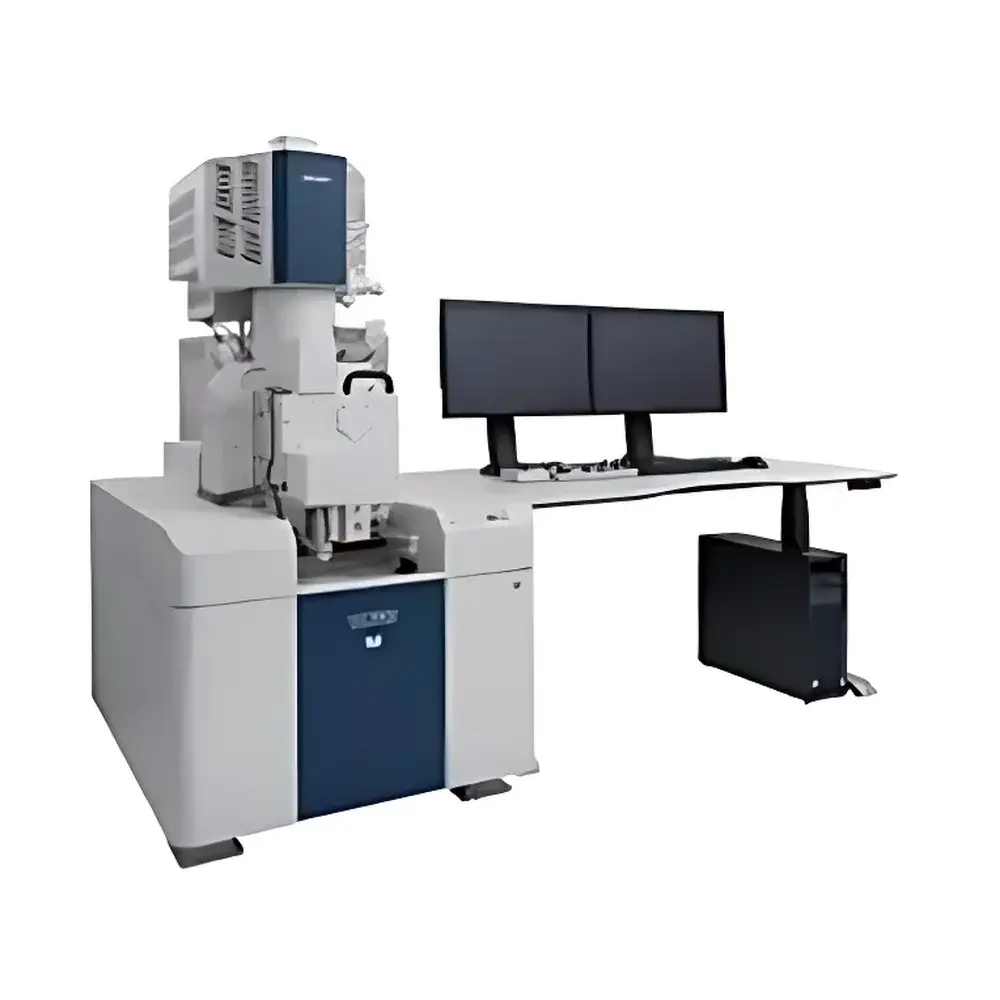
Hitachi SU7000 Cold Field-Emission Scanning Electron Microscope
| Brand | Hitachi |
|---|---|
| Origin | Japan |
| Manufacturer | Hitachi High-Tech Corporation |
| Product Type | Imported Instrument |
| Model | SU7000 |
| Electron Gun | Cold Field-Emission (CFEG) |
| Secondary Electron (SE) Resolution | 0.8 nm @ 15 kV, 0.9 nm @ 1 kV |
| Magnification Range | 20× to 2,000,000× |
| Accelerating Voltage | 0.1–30 kV |
| Backscattered Electron (BSE) Resolution | Not Specified |
| Detector Channels | Simultaneous 6-channel signal acquisition and display |
| Maximum Image Resolution | 10240 × 7680 pixels |
| Sample Chamber | Ultra-large chamber with 18 accessory ports |
| Vacuum Mode | High vacuum and low vacuum (down to 300 Pa, optional) |
Overview
The Hitachi SU7000 is a high-performance cold field-emission scanning electron microscope (CFEG-SEM) engineered for nanoscale imaging, compositional analysis, and structural characterization in advanced materials science, semiconductor metrology, life sciences, and failure analysis laboratories. Utilizing a high-brightness cold field-emission electron source, the SU7000 delivers exceptional beam coherence and stability—enabling sub-nanometer resolution imaging at low accelerating voltages without compromising signal-to-noise ratio. Its optical column integrates a patented in-lens secondary electron detector and multi-segment backscattered electron detector, optimized for simultaneous, spatially registered acquisition of topographic, compositional, and crystallographic contrast signals. Unlike conventional SEMs requiring iterative working distance (WD) adjustments to balance SE/BSE collection efficiency, the SU7000’s re-engineered sample chamber and detector geometry enable consistent, high-fidelity signal separation at a fixed WD—reducing setup time and enhancing measurement repeatability across multi-modal workflows.
Key Features
- Cold field-emission electron gun (CFEG) providing long-term emission stability, minimal energy spread (<0.3 eV), and high current density for high-resolution imaging and microanalysis.
- Simultaneous 6-channel signal acquisition system supporting real-time overlay and correlation of secondary electrons (SE), backscattered electrons (BSE), absorbed current, and energy-dispersive X-ray spectroscopy (EDS) signals.
- Ultra-large sample chamber accommodating specimens up to Ø300 mm × 100 mm height, with 18 standardized accessory ports for in-situ stages, cryo-transfer systems, EBSD detectors, and environmental SEM (ESEM) modules.
- Low-vacuum capability down to 300 Pa (optional configuration), enabling direct observation of non-conductive, hydrated, or beam-sensitive specimens without metal coating.
- High-definition imaging at up to 10240 × 7680 pixels (78.6 MP), with pixel dwell times configurable from 10 ns to 10 µs for optimized signal fidelity across dynamic and static imaging modes.
- Integrated lens aberration compensation and stigmator auto-tuning algorithms ensure consistent focus and astigmatism correction across the full magnification range (20×–2,000,000×).
Sample Compatibility & Compliance
The SU7000 supports diverse sample types—including bulk metals, ceramics, polymers, biological tissues, geological specimens, and nanomaterials—without mandatory conductive coating in low-vacuum mode. Its robust chamber design complies with ISO 14644-1 Class 5 cleanroom requirements for installation integrity and meets IEC 61000-6-3 electromagnetic compatibility standards. For regulated environments, the system supports audit-ready operation under GLP and GMP frameworks when paired with optional 21 CFR Part 11-compliant software modules, including electronic signatures, user access control, and immutable acquisition logs. All vacuum components are certified to ISO 2859-1 sampling plans for incoming inspection, and column bake-out protocols follow ASTM E1558 guidelines for residual gas analysis.
Software & Data Management
Hitachi’s proprietary “SmartSEM” platform provides unified control of imaging, stage navigation, signal processing, and analytical quantification. The software architecture supports DICOM-compatible export, TIFF/RAW binary output with embedded metadata (accelerating voltage, WD, dwell time, detector gain), and batch-processing pipelines for automated particle analysis, grain size distribution, and phase mapping. Data integrity is ensured via SHA-256 hash verification on acquired images and spectra; raw data files are stored in vendor-neutral HDF5 format to facilitate third-party integration with Python-based analysis tools (e.g., HyperSpy, scikit-image). Remote operation and collaborative annotation are enabled through TLS-encrypted web interface, compatible with institutional SSO authentication.
Applications
- High-resolution defect inspection in 3D NAND and GAA-FET semiconductor devices.
- In-situ tensile and thermal testing using integrated mechanical and heating stages.
- Correlative microscopy workflows combining SEM imaging with Raman spectroscopy and AFM topography.
- Quantitative EDS mapping of trace elements in battery cathode materials (e.g., Ni-rich NMC) at sub-10 nm spatial resolution.
- Morphological and elemental analysis of extracellular vesicles and freeze-fractured membrane structures in cryo-SEM mode.
- Automated mineral liberation analysis (MLA) in mining and metallurgical process optimization.
FAQ
What vacuum modes does the SU7000 support?
The SU7000 operates in high vacuum (≤1×10⁻⁴ Pa) by default and optionally supports low vacuum mode down to 300 Pa for uncoated, insulating, or hydrated samples.
Is the cold field-emission gun user-replaceable?
No—the CFEG is factory-aligned and sealed; replacement requires certified Hitachi service engineers and chamber venting/re-pumping procedures.
Does the system support automated EDS quantification per ASTM E1508?
Yes—SmartSEM includes ZAF matrix correction routines, standardless quantification, and drift-corrected spectral acquisition compliant with ASTM E1508 and ISO 16591.
Can the 6-channel display be customized per user role?
Yes—display layouts, detector assignments, and hotkey configurations are saved per user profile and enforce role-based permissions.
What is the maximum stage tilt angle with full detector clearance?
The motorized stage provides ±90° tilt with automatic detector retraction, maintaining optimal SE/BSE collection geometry across the full range.


